
Contents
Visual Field Test
Your visual field is how broad of an area your eye can view when you concentrate on a center point. The visual field test is one way your ophthalmologist estimates how much you’re able to see from one eye and how much vision loss has taken place over time.
A visual field test can discover if you have blind spots called scotoma in your vision and where they are exactly present in the eye.
A scotoma’s size and shape tell how eye disease or a brain disorder is harming your vision. For example, if you have glaucoma, this test helps to determine any feasible side (peripheral) vision loss from the disease.
Ophthalmologists also use a visual field test to evaluate how vision may be defined by eyelid problems like ptosis and droopy eyelids.
Your ophthalmologist may use data from the visual field test to find diseases like:
- Glaucoma
- Macular degeneration
- Optic glioma
- Brain tumor
- Multiple sclerosis
- Stroke
- Temporal arteritis
- Central nervous system disorders
- Pituitary gland disorders
- High blood pressure
- One can go for glaucoma surgery after having the test.
Uses of Visual Field Test
The visual field test is most commonly used to identify signs of glaucoma that harm the optic nerve. Moreover, a visual field test is useful for the disclosure of primary or peripheral retinal diseases of the retina, eyelid conditions like drooping, optic nerve damage, and occipital cortex.
The uses of visual field testing are:
- Used for visual eye testing in a screening of glaucoma. Peripheral vision loss is usually an initial and detailed sign of glaucoma.
- Used for screening and measuring for eyelid droop i.e ptosis.
- Testing toxicity from some medicines like screening the toxicity from hydroxychloroquine that can harm the central retina.
- Estimating the size of retinal diseases like retinitis pigmentosa, retinal detachment, etc.
- Identifying conditions that can change the visual pathways from the optic nerves to the occipital lobe of the brain, involving tumors, inflammatory disease, increased intracranial pressure, eye injury, etc.
- Examination for malingering behavior or mental disorders.
Visual Field Testing Procedure
There are a variety of techniques to estimate the visual fields. The visual field test is performed in one eye at a time, with another eye fully covered to avoid errors. In testing, the patient must look straight ahead at every time to correctly map the peripheral visual field. The most common visual field testing methods are used to observe fixation or the capacity of the patient to keep a constant straight-ahead gaze.
Types of Visual Field Tests
A variety of sensitive tests for estimating visual field test loss exist, including:
Automated Perimetry
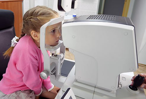
Different forms of automated perimetry tests contain your answers to the presence of objects in different areas of your field of seeing. While your head is retained still, normally with a forehead and chin stay inside a big bowl-like instrument. You look at a source of light straight ahead and tiny lights of different forces are flashed from random spots in your visual field.
Electroretinography
This test includes electrical activity produced by the photoreceptor cells in the retina. The eye is stimulated by a specific strobe light or a reversing checkerboard design of light. The measurement is seized by an electrode located on the cornea, and a graphic recorder named an electroretinogram is formed. Electroretinography helps diagnose many hereditary and acquired disorders of the retina.
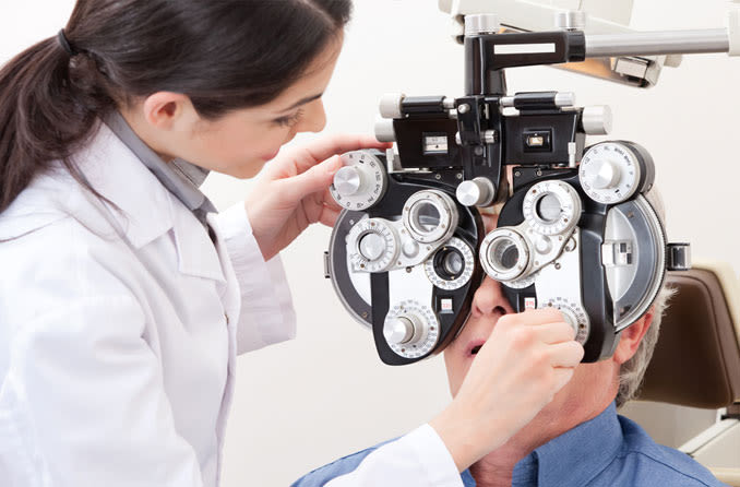
Confrontation Visual Field Testing
The doctor directs the patient to see straight ahead. The doctor will give stationary or moving targets in the patient’s peripheral visual fields. While keeping a straight-ahead gaze, the patient lets the doctor understand when he/she can view the target of the peripheral vision. The target may be a little disc on a stick but usually, the target is the doctor’s hand holding up 1 or 2 fingers.
Amsler Grid
This is a marked image of a grid with a dot in the center. The patient is directed to look at the dot, one eye at a time, and see whether the grid lines circling the dot look distorted, faded, or partly missing. This test is most often used to identify central visual field test defects.
Static Automated Perimetry
Pinpoint flashes of light of differing sizes and brightness are projected within a huge white bowl. The patient is claimed to look at the middle of the bowl and press a button each time light is observed in the peripheral vision. The machine receives the data and uses sophisticated software to examine the results.
Kinetic Perimetry
Moving targets of different light sizes and intensities are determined and the patient shows when they display visible in the peripheral vision. The resulting data is applied to map the full visual field. The full, normal range of the visual field increases about 120° vertically and almost 160° horizontally.
Frequency Doubling Perimetry
This test uses different intensities of a flickering image to examine the visual field. It is especially helpful in identifying early glaucoma field loss. Frequency doubling is based on an optical image produced with upright bars of varying colors normally black and white resembling a screen.
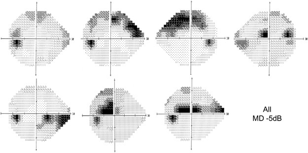
Visual Field Test In Glaucoma
Glaucoma is an eye disease that originally affects your side vision ( peripheral ). One of the key tests that your ophthalmologists will perform as part of the diagnosis for glaucoma is the visual field test, which is repeated annually to decide if the disease is permanent or becoming worse. There are many diverse types of visual field machines.
As the disease advances, more and more of the peripheral vision is lost until ultimately, in very late and high-level disease, the central vision is also impaired. Sometimes there are patients with glaucoma who have their central vision-impaired early in the way of the disease, which is a different reason that formal visual field testing is so valuable.
The repeated visual field test is a crucial part of building baseline visual fields and observing glaucoma over time. The test does have some variability, so repeating the test not only helps your eye doctor determine if a change is real but also will develop your test-taking ability over time.
It is helpful to understand that the test is designed to be challenging, so try not to be too stressed (you can take online stress counseling) while going for a visual test to resolve the problem of glaucoma. The test usually takes 5-10 minutes for each eye.
What Happens During The Visual Field Test?
A doctor will seat you conveniently in front of the machine and will use appropriate lenses to correct for any glasses correction you require. He or she will give you guidance on how to take the test. During the test, the specialist will examine to make sure that you are viewing straight ahead at the fixation light. Examine to make sure that your upper eyelid is large enough so as not to block your vision, and may kindly reposition you if your head has shifted too much.
Duration of Visual Field Test
The test, which includes the central and side vision for each eye, takes about 5-10 minutes, and you can blink commonly throughout. During the test, one eye is coated (so that one eye is tested at a time. And you want to constantly stare straight ahead at the calm yellow light. Then, other lights flash one at a time off to the side and you must press the button whenever you view one of these lights.
The test is created so that the light flashes slowly dim until you can no longer see them. So do not worry if it appears to be a long pause within flashes. You are not demanded to see all the lights, and indeed you may notice some of them. This also means that many of the lights you do view will be very low. Also, you can constantly pause the test by pressing the response button down if you need a rest. When you release the button the test resumes.
- Amsler grid and Confrontation visual field take a few minutes to be treated.
- A static field for lid droop or ptosis screening takes about 8 minutes.
- The static field for comprehensive glaucoma evaluation takes about 15 minutes.
- The Frequency doubling perimetry testing for glaucoma screening takes approximately 10 minutes.
- Kinetic Goldmann Perimetry for the entire glaucoma assessment is about 20 minutes procedure.
Monitoring the condition
After you have taken the test already, you will understand what to expect. And then the task of monitoring your glaucoma and making sure that it continues steady will start during your follow-up visits.
There are quite typical variations that doctors see in glaucoma, including the shape and place of any defects. Also, when there is more central vision loss or if you have a weaker vision in one eye, many algorithms can be done.
Sometimes, other tests will be used to observe changes in your vision, in addition to the visual field test, because they can give various types of helpful information.
It is very important to check the retina and optic disc correctly to evaluate whether or not a visual field defect meets the front of the disc and retina, or fits with other clinical signs.
Cost of Visual Field Test
The visual field test is performed most often by eye doctor’s optometrists, ophthalmologists, and neurologists. The cost of visual field testing is usually covered by health insurance. These visual fields test your side or peripheral vision. The process is fast and painless. The test costs $15 and Indian Rs. 1,500-2,000. The visual field test is a vital part of glaucoma diagnosis and is repeated regularly to determine if the disease is permanent or getting more critical. The visual field test is estimated as a functional test.
It also enables your doctor to tell you if you have lost any field of vision from glaucoma. Also, help to define the rate of disease progression. Your doctor will be capable to tell you how critical your disease is based on this test. Although the test is not painful, and it just takes a few minutes for each eye. Many eye and brain diseases can cause peripheral vision loss and other visual field irregularities. The visual field test is done by eye care professionals to recognize blind spots and other visual field defects, which can be an early indication of these problems.
For regular eye checkups and visual field test, do visit our website Eyemantra.
To get your regular eye checkups thoroughly from an expert ophthalmologist, you can book appointments at +919711115191. Or mail at [email protected].
We also offer various services like Retina Surgery, Specs Removal, Cataract Surgery, and much more.
Medically Reviewed By

Senior Eye Surgeon | LASIK, SMILE & Cataract Specialist