

Contents
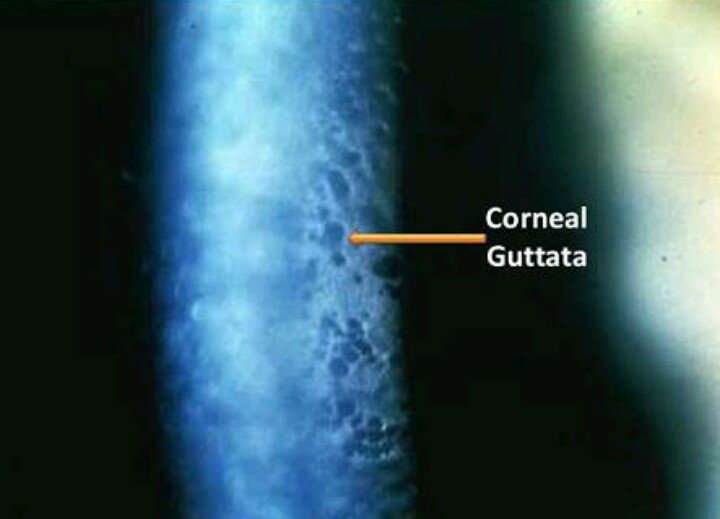
What Is Cornea Guttata?
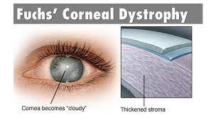
Cornea guttata is a degenerative disease. It causes the accumulation of focal outgrowths in form of droplet-shaped bulges, by the corneal surface, which is the early stage of the disease. Corneal guttata is an initial stage of “Fuchs’ dystrophy” that occurs in several stages.
Patients who are suffering from cornea guttata disease have a huge risk of vision loss when they have corneal transplantation. Cornea guttata is a disease in which the patients have droplet-shaped bulges over the corneal surface inside the eye. There is an accumulation of focal outgrowths which are called ‘guttae’ in cornea guttata.
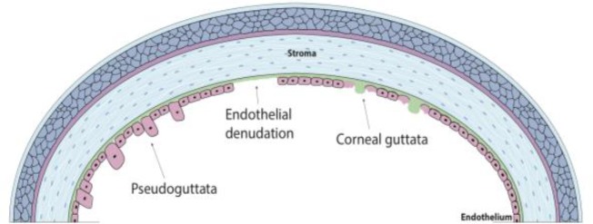
Corneal epithelium cells are used in corneal transplant surgery. It causes problems in the corneal epithelium leading to surgery complications. In cornea guttata, the membrane gets thickened by the increase in collagen accumulation and a number of guttae. The decrease in endothelial cells causes endothelial edema. i.e, inflammation or swelling which causes an influx of aqueous humor into the cornea. This results in blurred vision or severe cases of loss of vision.
Symptoms of Cornea Guttata
This disease is an alternative to a corneal disease which is called Fuchs’ dystrophy. It causes loss of vision or impairment of vision, as it affects the cornea of the eye. A person with this disease can face problems such as-
- loss of transparency inside the eye
- Fluctuating and reduced vision
- Thickening of cornea
- Severe pain
- Compromised vision in severe cases.
- No perception of images, during morning
- Loss of acuity
Risk Factors of Cornea Guttata
The cornea guttata conditions are usually seen in people who are aged above 40 years. The condition in people who are young is rarely seen i.e, between 30 to 40 years of age. Women are easily affected by this condition.
The person suffering from cornea guttata condition usually tends to pass on their disease through genetic factors i.e., have 50% chances to pass on their gene to their progeny. Usually, this condition is seen in women.
Diagnosis of Cornea Guttata
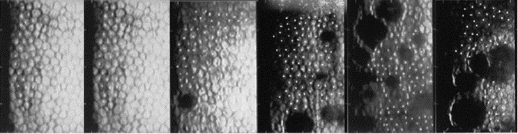
An eye examination using a slit lamp helps in the identification of cornea guttata condition. As the droplet-shaped bulges are seen on the cornea. Usually, in the central cornea, the guttata are seen and present in both eyes. Though, the guttata present in one eye can be more in number and severe than in the other eye. The increase in severity in cornea guttata condition leads to the development of haze in the corneal stroma.
As this stroma state increases, it thickens the stoma and causes visibility of endothelial cells of the cornea, and even more, cells get damaged.
Clinical Diagnosis
By use of pachymetry i.e., the measurement of the corneal thickness of the eye is done. As the disease increases, the corneal thickness increases in size. So, the measurement of corneal thickness helps in diagnosing the disease. The measurement of corneal thickness has some other advantages i.e., it also helps in the analysis of risk/benefit ratio in other surgeries such as cataract surgery.
The changes in endothelium and mild corneal stromal edema helps in the early diagnosis of cornea guttata cases. The patients can be guided upon the severity of the corneal guttata condition, by understanding the endothelial changes and their cell count. The endothelial changes can be diagnosed with the help of specular microscopy.
Surgical Procedure of Guttata
Various surgical procedures are used for treating this condition when the medical procedures cannot help in treating cornea guttata. The procedures are
- Penetrating keratoplasty
- Descemet’s stripping endothelial keratoplasty
- Descemet’s membrane endothelial keratoplasty
- Descemetorhexis Without Endothelial Keratoplasty
After Guttata Surgery
The patient is asked to come more often to check up after a few weeks, after the surgery, so that the doctor can check for any kind of problems inside the eye. Evaluation of transplant health wound healing and recovery of vision is checked thorough checkups. Not only have that, even the removal of sutures reducing astigmatism.
Surgical procedures that include the transplantation process have more potency for rejection than normal organ transplant surgery. At any point of the surgery, the cornea transplant can be rejected by the eye. So, a check-up is required for the treatment of any kind of rejection of the transplant. Glaucoma can be seen after surgery in some patients as an adverse effect. Glaucoma can cause permanent blindness in the patient.
Complications Due To Surgical Procedure
The complications which are seen in the surgical procedure are infection, bleeding and wound leakages, and problems caused due to sutures.
The use of typical steroids for the long term can cause other eye-related complications such as cataracts and glaucoma inside the eye. Sometimes due to the surgeries, there is a risk of detachment of graft with a need of re-bubbling constantly. loss of vision and severe eye pain can be caused due to severe complications.
Relation of Cornea Guttata To Phaco
After the phacoemulsification method, the patients who are suffering from cornea guttata have a major risk for a corneal transplant. Cornea guttata condition causes the damage of endothelial cells. So, the patients who are suffering from cornea guttata do not undergo the transplant because it increases the loss of cells and increases in Cornea guttata condition even more.
Fuchs’ endothelial corneal dystrophy also known as FECD, is a disease whose initial stage is cornea guttata and it is a bilateral degenerative corneal disease which in severe cases causes an increase in corneal swelling and endothelial decomposition occurs, this results in impairment of vision. Cornea guttata is just the early stage of Fuchs’ dystrophy, but this condition can also be seen in glaucoma, inflammation, traumatic conditions, and aging.
The phacoemulsification in patients, who are suffering from cornea guttata is correlated with corneal transplantation. In the first year after phacoemulsification, the patients have reduced in number. An increase in age during phacoemulsification causes a decrease in the probability of patients who are undergoing corneal transplant surgery.
Treatment of Cornea Guttata
Some patients who are suffering from the disease and mild guttata condition, and have no corneal stromal edema need to go for a check-up every 6 to 12 months. A patient who is suffering from a severe case must be checked more often so that the treatment is adequate. Patients who are using bandage contact lenses have more risk of getting an infection, so they must check even more.
It can be removed by the use of eye drops and hypertonic saline or ointments. The hypertonic saline is used for removing the excess water from the bulges present over the cornea. The decrease in water from the cornea helps in reducing cornea guttata and decreases the time taken for recovery of vision.
Various activities help in the recovery of vision. Some are the usage of hair dryers to provide clear vision by blowing air. In severe cases, the use of bandage contact lenses can be helpful in treatment. Surgery can be the last option for treatment when the cornea guttata condition becomes more severe i.e., Fuchs’ dystrophy.
Since transplantation deals with a much more complex post-operative procedure as it lasts for life long. So, the study of patients who are suffering from this condition and want to have corneal transplantation surgery during their initial years of cataract surgery would be really interesting.
Visit our website Eyemantra
To book an appointment call +91-9711115191. Or mail us at [email protected]. Our other services include Retina Surgery, Specs Removal, Cataract Surgery, and many more.
Medically Reviewed By

Senior Eye Surgeon | LASIK, SMILE & Cataract Specialist