

Contents
Slit Lamp Examination
The slit lamp examination is a technique that is used for diagnosing the eye. The examination includes testing the functioning as well as the presence of any problem in the eye.
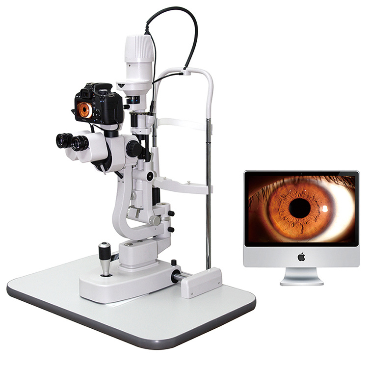
The slit lamp is a microscope that is used for diagnosing of eye-related diseases as well as to check the functioning of the parts of the eye which cannot be seen with normal technique. This microscope uses bright light which allows it to view all the structures which are present inside the eye. This equipment allows the ophthalmologist to have a closer look at the structures inside the eye.
This slit lamp examination is also called biomicroscopy. It allows the ophthalmologist to view the functioning as well as the presence of any kind of abnormalities. Any kind of inflammation or injury of the various structures inside the eye at the microscopic level is also tested.
Preparation for The Slit Lamp Exam
The person who is taking this exam does not require any kind of special preparation before the exam. But, the dilation of the pupil is done with the help of dilating drops that are dropped inside the eye. This dilation can stay for few hours after the examination is completed.
So, the patient must not drive the car or any kind of vehicle immediately after the slit lamp exam. Not only this but after dilation the vision of the patient is blurry. And there is an increased sensitivity towards the light for several hours post-slit-lamp examination. So, the patient must carry sunglasses to prevent this sensitivity. And tag along a person to look after the patient, post-slit-lamp examination.
Slit-lamp Examination Procedure
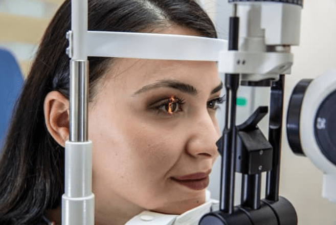
In the slit lamp examination, the person is made to sit on a chair. An instrument is placed over which the person is asked to rest his/her chin as well as forehead. This helps in maintaining your head steady and straight while performing the slit lamp examination.
The doctor puts drops inside the eye so that the abnormalities which are present on the corneal surface or inside the eye can be seen clearly. And any kind of eye disease can be diagnosed properly. These drops are composed of fluorescein which is a dye that is yellow in color. Other drops that cause dilation of a pupil are added so that the slit lamp examination can be done properly.
Along with the slit lamp microscope which has a high-intensity light, a low-powered microscope is also used in the slit lamp examination. This slit lamp microscope has different kinds of filters that help in viewing different structures present inside the eye. The changes inside the eye over time can be tracked with the help of digital images, which are captured with devices that are used at some hospitals.
The doctor passes a high-intensity light inside the eye through the microscope. This helps in viewing the different structures inside the eye. This high-intensity light does not cause any damage to the eye.
Diagnosis Of Different Eye Part With Slit Lamp
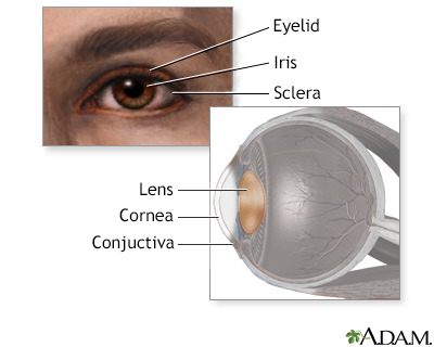
The slit lamp examination is used for viewing various structures of the eye such as eyelids, conjunctiva, cornea, retina, and optic nerve. Firstly, the ophthalmologist performs the test at the front side of the eye. And then he performs the slit lamp test on the backside of the eye. At the front side the doctor checks for:
Sclera
It is made up of tough, fibrous tissue that makes up the outer protective layer of the eye. It is white in color. This exam is used for detecting any kind of inflammation or discoloration of the sclera. As it indicates the presence of eye diseases such as scleritis or any autoimmune disease which can cause permanent vision loss of the patient.
The examination can help in diagnosing the presence of conjunctivitis or pink eye condition which is caused by an infection or an allergy of conjunctiva i.e, is a thin and transparent layer that is covered over the sclera.
Cornea
It is a dome-shaped structure that is present in the front part of the eye. The slit lamp examination can help diagnose the dry eye condition. Dry eye is caused by abnormal functioning of the tear film of the eye and blockages inside the tear gland that can provide enough moisture to the eye.
The slit lamp examination can detect any kind of abnormal structures over the corneal surface which can be a sign or symptom for any kind of corneal disease such as corneal dystrophy which causes blurry vision and permanent loss of vision if left untreated.
The use of fluorescein dye helps in highlighting some of the structures and identifying any kind of injuries present inside the eye near the cornea surface such as corneal abrasion or any kind of infections such as herpes keratitis.
Lens
The slit lamp examination is used for diagnosing cataract which is a disease in which the clear part of the eye i.e, the lens becomes cloudy and becomes difficult to view through the cloudy lens and decreases the vision of the eye. The lens focusses on the light over the retina so that the image is formed clearly.
The cataract condition is common in the case of aging process and as the cataract condition becomes more severe it needs to be removed with the help of a surgery.
This slit lamp microscope is also used for giving a detailed and closer look of the structures that are present at the back side of the eye. These structures can be seen with the help of dilating drops which are put inside the eye. The structures which can be seen at the back side of the eye are:
Retina
It is a layer of nerve cells which are lined against the back wall inside the eye. This structure absorbs light and helps in formation of images which can be easily understood by the brain via optic nerve. This slit lamp microscope helps in diagnosing various problems that are caused to the retina such as tearing of the retina or detachment of the retina. These conditions can cause the permanent loss of vision. This slit lamp microscope can also diagnose various disease such as presence of an infection or any kind of injury and also degeneration of the blood vessels inside the eye. This slit lamp can also detect the increase in the pressure inside the eye i.e, intraocular pressure.
For example, in the case of age-related macular degeneration (AMD), there are yellow deposits that are seen in the macular region. This disease affects the central vision of the eye. This can be detected with the help of a slit lamp microscope. The slit lamp microscope also detects inherited diseases such as retinitis pigmentosa.
Optic nerve
This nerve acts as a connection between the brain and the eye. It is located at the back of the eye. The slit lamp microscope helps in diagnosing the glaucoma condition which if is not treated in early time can cause permanent loss of vision and this glaucoma condition mostly affects the optic nerve which causes loss of vision.
Eye Conditions This Exam Help Diagnose?
A slit lamp exam can help detect the following eye conditions:
- Macular Degeneration: A chronic age-related condition that affects the part of the eye that is responsible for central vision.
- Detached Retina: An eye condition when the retina, the inner layer of tissue at the back of the eye, becomes detached from its base. Having a healthy retina is important for good vision.
- Cataracts: A clouding of the lens that negatively affects the ability to see images clearly. A cataract is also an age-related condition.
- Injured Cornea: The cornea, one of the tissues that cover the surface of the eye. It protects the eye from harmful particles. The slit-lamp exam can detect the cornea’s condition.
- Blockages of the retinal vessel: obstructions in the eye’s blood vessels that can cause a sudden or gradual vision loss.
The best way to treat your eyes is to visit your eye care professional and get your eyes checked regularly. He will be able to assess the best method of treatment for your eye ailment.Visit our website Eyemantra.To book an appointment call +91-9711115191. Or mail us at [email protected]. Our other services include Retina Surgery, Specs Removal, Cataract Surgery, and many more.
Related Articles :
Medically Reviewed By

Senior Eye Surgeon | LASIK, SMILE & Cataract Specialist