
Imagine a life where your vision is sharp, clear, and vibrant. But for many, this seems like a distant dream, especially if you’re dealing with corneal issues. However, here’s the good news: there’s a solution, and it starts with understanding the essential tests before a corneal transplant.
In this blog, we’re going to walk you through the critical examinations that pave the way for a successful corneal transplant surgery. By the end of this journey, you’ll not only know what to expect but also feel confident and empowered about taking the next step towards clear vision. So, let’s dive into the vital tests that lay the foundation for a successful corneal transplant!
Contents
- 1 Key Tests Required To Under Before a Corneal Transplant
- 1.1 Assessing Your Vision: The First Step in Pre-Transplant Testing
- 1.2 The Slit Lamp Exam: A Detailed Eye Examination for Corneal Health
- 1.3 Dilated Eye Exam: Unveiling Retinal Health in the Pre-Transplant Process
- 1.4 B-Scan: Testing White Cornea to Study Internal Eye Structures
- 1.5 Specular Microscopy: Examining the Endothelium, the Final Corneal Layer
- 2 Conclusion
Key Tests Required To Under Before a Corneal Transplant
Assessing Your Vision: The First Step in Pre-Transplant Testing
 Before the journey to a successful corneal transplant surgery begins, it’s crucial to lay the groundwork by assessing your vision. This first step in the pre-transplant testing process aims to evaluate your current visual condition and determine your prescription glasses number. Here’s what you need to know:
- Visual Acuity Test
This test measures how well you can see at different distances. You’ll be asked to read letters or symbols from a standardized chart, with one eye covered at a time. The results help determine your level of visual impairment. - Refraction Test
During this test, an optometrist or ophthalmologist will use a phoropter or trial frame to determine your exact eyeglass prescription. By assessing how lenses affect your vision, they can identify the right prescription to correct your refractive errors, such as myopia (nearsightedness) or hyperopia (farsightedness). - Cycloplegic Refraction
In some cases, a cycloplegic refraction test may be performed, which involves using eye drops to temporarily paralyze the muscles that control the shape of the eye’s lens. This test provides an even more accurate prescription, especially for individuals with high refractive errors.
The results of these tests offer a clear picture of your current vision and the degree of correction needed. This information is essential for planning your corneal transplant procedure and ensuring the best possible outcomes.
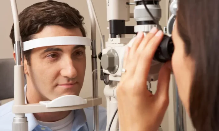
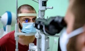
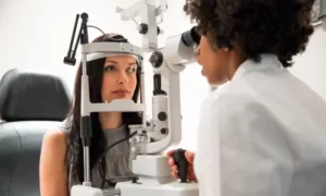
The Slit Lamp Exam: A Detailed Eye Examination for Corneal Health
 The journey towards a successful corneal transplant continues with the second crucial step: the slit lamp exam.
Why It Matters: The condition of your cornea plays a vital role in the success of the corneal transplant procedure. For this, a thorough slit lamp examination provides valuable insights that guide your surgeon in planning the surgery and addressing any underlying corneal issues. It’s an essential step in your journey to clear vision and an improved quality of life. Let’s dive into the details of this essential test:
Understanding the Slit Lamp Exam: The slit lamp exam, also known as biomicroscopy, employs a specialized microscope called a slit lamp. This instrument allows your ophthalmologist to examine the different layers of your cornea with exceptional precision. Here’s how the process unfolds:
- Patient Positioning
You’ll be seated comfortably, and the slit lamp will be positioned in front of your eye. - Focused Light
The slit lamp emits a narrow, intense beam of light, which is directed onto your cornea. This focused light allows the examiner to see the corneal details more clearly. - Ocular Examination
Using high magnification and various lenses, the ophthalmologist will carefully examine the entire cornea. They’ll assess its transparency, look for any irregularities, and check for conditions such as scars, dystrophies, or infections. - Assessing Corneal Health
The slit lamp exam is a critical part of the pre-transplant testing process. It helps identify any corneal issues that need to be addressed before the transplant, ensuring the health and integrity of the cornea.
Dilated Eye Exam: Unveiling Retinal Health in the Pre-Transplant Process
 As we move forward in the pre-transplant testing process, we arrive at the third essential step: the dilated eye exam. This examination is crucial for checking the health of your retina, uncovering any potential eye issues that might impact the success of your corneal transplant. Here’s what you need to know:
- Dilation: You’ll receive eye drops that contain medications to dilate your pupils. It takes approximately 20-30 minutes for the drops to take effect.
- Pupil Enlargement
Once your pupils are sufficiently dilated, your eye care provider will use a special magnifying lens and a bright light to examine the interior of your eye. - Retinal Examination
The focus of this examination is your retina. Your eye care provider will look for any signs of retinal diseases or conditions that might affect your vision and the success of your corneal transplant. - Assessing Blood Vessels
The dilated eye exam also allows for a close examination of the blood vessels in your eye, which can reveal issues such as diabetic retinopathy or age-related macular degeneration.
The Importance of Retinal Health
A healthy retina is essential for clear vision and the success of your corneal transplant. This examination helps identify any retinal problems that need to be addressed before the transplant, ensuring that your eye is in optimal condition for the procedure.
B-Scan: Testing White Cornea to Study Internal Eye Structures
 In some cases where the white cornea obstructs the view of the retina, a specialized test known as the B-scan becomes essential. Let’s explore the significance of the B-scan:
Understanding the B-Scan: The B-scan, short for brightness scan, is an ultrasound test that utilizes high-frequency sound waves to create images of the eye’s interior. It’s a valuable tool when the cornea’s cloudiness or opacity prevents a direct view of the retina. Here’s how the B-scan procedure works:
- Patient Preparation
You’ll be comfortably positioned, and a small amount of gel is applied to your closed eyelid. - Transducer Probe
A transducer probe, which emits and receives sound waves, is gently placed on the gel-covered eyelid. - Sound Wave Emission
The probe sends out high-frequency sound waves that bounce off the internal eye structures, creating detailed images. - Image Display
The captured images are displayed on a screen, allowing the eye care provider to assess the condition of the retina, vitreous, and other internal eye components.
The Role of the B-Scan
The B-scan is a crucial test when a cloudy cornea obscures the view of the retina. By providing images of the internal eye structures, it helps the medical team identify any abnormalities, such as retinal detachments, vitreous hemorrhages, or tumors. These insights are valuable for planning the corneal transplant surgery and ensuring the overall health of your eye.
Specular Microscopy: Examining the Endothelium, the Final Corneal Layer
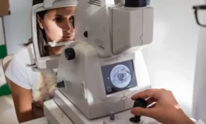 In the final step of our pre-transplant testing journey, we delve into specular microscopy—a vital examination of the endothelium, the last layer of your cornea. This test ensures the health and integrity of this crucial corneal component. Let’s explore the significance of specular microscopy:
Unpacking Specular Microscopy
Specular microscopy is a non-invasive imaging technique that focuses on the endothelium—a single layer of cells that lines the inner surface of your cornea. This examination uses a specialized microscope equipped with a camera to capture images of these cells. Here’s how it works:
- Patient Positioning
You’ll be seated comfortably, and your eye will be positioned in front of the specular microscope. - Image Capture
The microscope emits a thin beam of light onto the cornea’s surface, and the camera captures high-resolution images of the endothelial cells. - Analysis
The obtained images are carefully examined to assess the density, shape, and overall health of the endothelial cells.
By undergoing specular microscopy, you’re safeguarding the health and integrity of the endothelium, the final layer of your cornea. This step is pivotal in preparing for a successful corneal transplant, ensuring the best possible outcome and a brighter, clearer future.
Conclusion
In your journey towards a brighter and clearer vision, you’ve taken a significant step by exploring the essential tests required before a corneal transplant. These tests are the foundation on which successful corneal transplant surgeries are built. They provide a comprehensive understanding of your eye’s condition, ensuring that the transplant procedure can proceed smoothly and with the best possible outcome.
If you’ve been living with blurred vision, discomfort, and limitations due to corneal issues, the next step is clear. It’s time to take action. If you’re experiencing cornea-related problems, a corneal transplant surgery at EyeMantra can help you regain a clear vision.
Book your free appointment now at 9711116605. The team at EyeMantra is here to guide you, support you, and help you embark on a journey to a world seen through crystal-clear eyes. Your path to a brighter vision begins here.
Medically Reviewed By

Senior Eye Surgeon | LASIK, SMILE & Cataract Specialist