
The eye is the first amongst our five senses to be treasured. It is also the smallest and the most complex organ in our body. With its maximum diameter just about 2 cms, generally. The anatomy of the eye includes 2 million moving parts. which is just next to that of the brain. They can distinguish between more than 500 shades of any single colour and see more than 2.7 million colours.
How does The Eye work?
Sight is all about light. Light reflects from an object in the field of our vision. And it enters our eye through the cornea lens (the eye’s front window, the transparent outer covering of the eye). Just behind the thin veil of tears in the front. Passing through this clear layer helps focus the light.
Next is another layer of liquid called aqueous humour. Its purpose is to circulate throughout the front part of the eye and keep the inside pressure constant. After the aqueous humour, light moves through the iris. This is a coloured ring-shaped membrane. It has an adjustable circular opening called the pupil which dilates or compresses to control the amount of light coming in.
Now comes the lens. It operates just like a camera to focus light. It adjusts shape depending on whether the light is reflecting off an object near to you or far away.
The interior part of your eyeball is filled with a gel-like mass called vitreous humour. After passing through the lens, light has to travel through this humour.
Finally, it hits the sensitive layer of cells called the retina. At the back, it is “encoded” by a light-sensitive membrane called the retina. The cells are called photoreceptors. The retina transforms the image into electrical messages. as electrical impulses to our brain. Turns them into electrical impulses. These impulses are sent to the optic disk on the retina where they get transferred by a further set of electrical impulses along the optic nerve. And sent to the brain to be processed.
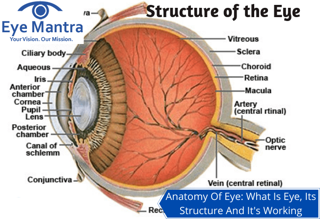
Anatomy of The Eye
| Part of the Eye | Primary Function | Simple Analogy |
| Cornea | The clear outer layer that focuses incoming light. | Protects the inside and provides a clear view. |
| Sclera | The tough white outer coat that maintains the eye’s shape. | A protective casing for the delicate inner parts. |
| Iris | The colored part that shrinks or expands to control light. | Adjusts the opening based on brightness. |
| Pupil | The black opening in the center that lets light in. | The hole through which light enters the camera. |
| Lens | Changes shape to fine-tune focus for near or far objects. | Zooms in or out to keep the picture sharp. |
| Retina | Layers of tissue at the back that sense light and color. | Captures the image to be processed. |
| Macula | A small, specialized area of the retina for sharp, central vision. | The part that handles the fine details. |
| Optic Nerve | Transmits electrical impulses from the eye to the brain. | Plugs the camera into the computer (the brain). |
| Vitreous Humor | A clear, jelly-like substance that fills the space behind the lens. | Keeps the eye inflated and round. |
| Ciliary Muscles | Muscles that pull on the lens to change its focus. | Does the work to keep images clear. |
The Outer Layer (Protection)
Sclera
Sclera is the tough skin that covers the outside of the eyeball. Though, it doesn’t cover the transparent Cornea. A protective coat on the “white” part in the anatomy of the eye.
Cornea
This is the transparent skin that covers the front of your eye. It is clear and slightly convex. This is the see-through part of the eyeball. It has no blood vessels in it.
Conjunctiva
This is the wall on the inside of your eyelid and the outside of the front of your eye (except for the special skin of the Cornea). You can also watch some tiny blood vessels on the Conjunctiva over the anatomy of your eye. Whenever your eyes get sore, these blood vessels get bigger, making your eye looks red.
Tear glands
These are small glands inside the upper eyelid. Their purpose is to make tears to keep the surface of the eyeball clean and moist. It helps protect our eyes from damage. When we blink, the tears cover the surface of the eye. Tiny particles that may be on our eye (like specks of dust) get washed into the corner of our eyes next to our nose. Sometimes, the tears may flow over our lower eyelids (when we cry or have a fever). But mostly the tears flow down a tiny tube at the edge of your lower eyelid, called the “Tear Ducts”, next to your nose. The beginning of that tube is visible as a tiny dot. This tube transfers the excess of tears to the back of our nose. This is why our nose “runs” when we cry.
The Middle Layer (Light Control)
Iris
Our Iris controls what amount of light would enter our eye. The Iris is the dark-coloured part of the anatomy of our eye. It consists of thin circular and longitudinal muscle fibres just behind the cornea. It forms a coloured muscular diaphragm across the front of our lens, with an aperture in the centre called the Pupil. This expands or contracts to allow more or less light, respectively, depending on the light in the surroundings. The layer of Aqueous Humour prevents the Iris from sticking to the lens behind and the cornea in front.
Pupil
This is the hole in the centre of the Iris. It lets light into our eyes. It shrinks in bright light and expands in dull light.
Lens
The lens is a clear crystalline globe, that focuses light onto the retina. It almost touches the posterior surface of the pupillary opening. Its shape is constantly modified to ensure that the ‘picture’ on the retina is as clear as possible. The Ciliary Muscles, attached to the surface of the lens, help the lens to change shape to focus. As the muscles contract, they cause the lens to become more round or long, so that the rays bend more or less, as per requirement.
If the object is far away, the Lens needs to bend the light rays from it more sharply, to make them fall on the centre of the retina, where vision is sharpest. For closer objects, the Lens becomes elongated so that light rays are bent less.
Ciliary Body
The Ciliary Body connects the Choroid to the Iris.
Choroid
The Choroid is the middle layer in the anatomy of the eye between the retina and the Sclera, which is separate from the RPE. It is made up of a lot of fine blood vessels that supply nutrition to the retina and the RPE. Moreover, it also holds a pigment that absorbs excess light to prevent blurred vision.
Ciliary Muscles
These are tiny muscles surrounding the lens. These muscles hold the lens in place. But they also play an important role in how we see them. They squeeze or relax to change the shape of the lens. They squeeze and contract, making the lens fat, to be able to look at nearby objects. And they relax, making the lens thinner, for faraway objects.
The Inner Layer (The Processor)
Retina
The working of the retina is much the same as a film in a camera. This layer is sensitive to light, lining the interior of the anatomy of our eye. It is made up of light-sensitive cells known as Rods and Cones. The human eye contains approximately 125 million Rods, which are necessary for seeing in dim surroundings. Whereas, Cones function best in bright light. We have about 6 to 7 million of them in the anatomy of our eye. They are indispensable for receiving a sharp accurate image. Cones can also distinguish colours. They turn the picture they receive into an electrical massage for the brain, which in turn translates that image to us.
Rod Cells
The other one of the 2 types of light-sensitive cells in the retina. There are about 125 million Rods, which are crucial for viewing in dim light. They contain a pigment “Rhodopsin” (Visual Purple) which is broken down in the light and regenerated in the dark. Breakdown of the Visual Purple gives rise to nerve impulses when all the pigment is bleached (i.e. in bright light) and the Rods no longer function. This is the condition when the cones get activated.
Cone cells
It is one of the two types of cells in the retina, that is light-sensitive. The human retina contains 6-7 million Cones. They operate best in bright light. They are quite essential for acute vision (receiving a sharp accurate image). The area of the Retina called the “Fovea” holds the most concentration of cones. Three types of cones are known. Each of them is sensitive to the wavelength of a different primary colour – red green or blue. Other colours are seen as a combination of these primary colours.
Macula
This is the yellow spot on the retina at the back of the anatomy of the eye. It surrounds the Fovea, the area that has the greatest concentration of Cone cells, and is, therefore, the area of greatest acuity of vision. When the eye is directed at an object, the part of the image that is focused on the Fovea is the image most accurately registered by the brain. It is situated right at the centre of our Retina. Because it’s the focal point of your eye, it has the most special, light-sensitive nerve endings, called Photoreceptors, than any other part.
Fovea
It is a small indentation at the centre of the Macula. The Fovea is described as the area with the greatest concentration of Cone cells, the messages encoded at the centre of the Fovea will be interpreted by the brain in the form of a visual image.
Retinal Pigment Epithelium (RPE)
It is a layer of dark tissue underneath the Photoreceptors. The purpose of these cells is to absorb excess light so the Photoreceptors can give a clearer signal. They also move nutrients to (and residual or waste from) the Photoreceptors to the Choroid.
Photoreceptors
They are of two kinds: rods and cones. They’re special nerve ends that convert the light into electrochemical signals.
Blindspot
Also called “scotoma”, a blind spot is a tiny part of the retina that is not sensitive to light. It is a small portion that has a close relation to the optic disk or optic nerve inside the retina. the reason for its presence in the ye is that it helps in carrying the images to the brain, where the image is processed.
The Connection to the Brain
Optic Disk
The visible portion of the Optic Nerve is also found in the retina. The Optic Disk identifies the start of the Optic Nerves where messages from Cone and Rod cells start from the eye through nerve fibres to the optic centre of the brain. This area is also known as the ‘blind spot’.
Optic Nerve
Optic Nerve is a continuation of the retina. Starting from the eye at the Optic Disk, it transfers all the visual information it gets to the brain, with the help of millions of nerve fibres branching from the Rods and Cones. It’s similar to the cable that carries all the TV pictures from your aerial to your TV so that you can watch the programs.
Internal Fluids
Aqueous Humour
Aqueous means related to water and humour is fluid. It is a water-like substance with certain compositions, and that flows through, also fills both anterior and posterior chambers of the eye.
The first and foremost role performed by the fluid (aqueous humour) is that it provides the eye tissues with nutrients to the avascular structure in the eye. So it brings glucose, amino acids as nutritive substances, and even ascorbic acid as an antioxidant, and some amount of oxygen as well. Those eye tissues which are aided by aqueous humour are namely, cornea and lens.
Vitreous Humour
This is a thicker liquid, gel-like, that fills the larger part of the eyeball and keeps it in shape. Vitreous means glassy, hence the name, because the vitreous humour is very clear so that light can pass through it.
Blood vessels
These are present all through the anatomy of the eye. And their function is to serve to bring oxygen and nutrients to the nerve cells.
Path of Vision
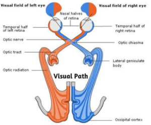
When the light rays fall on the Photoreceptor cells, changes occur in the pigments they contain. This leads to the bleaching of the pigments. And, thus, electrical impulses are generated. These get transmitted through a chain of neurons to the ganglion cells which carry the electrical impulses to the visual cortex of the brain. There they are processed and this is how we see an object.
Each eye receives data from half of the visual field. But the middle parts of both fields overlap slightly. This leads to Binocular Vision. However, the difference in the peripheral portions of the left and right fields of vision leads to the perception of Depth and 3-dimensional vision. It helps in assessing distances accurately and evaluating the depths and dimensions of objects.
Vision Problems/Diseases
The eyes are one of the most complex organs of the body. Thus, due to its complexity, it tends to be more fragile and vulnerable. Any carelessness can lead you to the following eye problems:
Nearsightedness (Myopia)
Myopia is a common vision problem in which you cannot see objects far away clearly. They appear to be blurry but can view nearby objects. It happens when the shape of your eye makes light rays bend (refract) wrongly, focusing images in front of your retina rather than on your retina. This can happen at any age and it may develop slowly or rapidly.
Nearsightedness usually names Nearsightedness. It usually occurs at the age of 40. But nowadays usage of laptops, mobile phones give a larger impact on the eye creating this problem. A basic eye exam can validate nearsightedness. You can pay for the blur with eyeglasses, contact lenses, or refractive surgery.
Farsightedness (Hyperopia)
There is a situation when we can see far objects but, when we got closer to that object everything seems blurry. It simply means an eye focus on faraway objects better than close objects. It’s the opposite of myopia, where you can see nearby objects but cannot see faraway objects.
The quality of your farsightedness defines your focusing capability. People with critical farsightedness may see only objects a large distance away. While those with mild farsightedness can see near objects. This problem can be corrected with eyeglasses, contact lens and the last option is surgery.
Astigmatism
Astigmatism is an error developed in the shape of the cornea/lens or just a slight imperfection in its shape. Usually, the cornea and lens shape are curved and are spread equally in all directions and are generally smooth. This greatly helps to converge the light rays hitting the cornea and lens to focus them onto the retina which is located at the back of the eye. If the cornea or lens shape is slightly irregular and isn’t smooth then it will not be bent properly and in doctor’s terms, it’s known as refractive error. This causes people to have blurry, hazy, or distorted vision.
The symptoms of astigmatism include haziness or blurriness in the vision, strain in the eye, frequent headaches, difficulty seeing at night, squinting while trying to see clearly, and discomfort in the eye.
Presbyopia
Presbyopia is the common loss of near-focusing ability that occurs with age. Most people notice the effects of presbyopia after age 40 when they start having difficulty seeing the small print.
Presbyopia happens naturally in people as they age. It may also be caused because of a lack of eye nutrition. The eye is not able to focus light directly on the retina due to the hardening of the natural lens. Ageing also breaks muscle fibres around the lens making it more difficult for the eye to concentrate on up-close things. The ineffective lens causes light to concentrate behind the retina, causing blurred vision for objects that are up close.
Cataracts
The leading causes of blindness, in the world, includes Cataracts (clouding of the lens). A cataract is known as the clouding of the lens in the eye which leads to a decrease in vision. Cataracts usually develop slowly over time and can affect one or both eyes. The symptoms of cataracts could be faded colours, blurred vision or double vision, halos around light, trouble with bright lights, and trouble seeing at night. This may result in having trouble driving, reading, or recognizing faces. Poor vision caused due to cataracts also results in an increased risk of falling and depression.
Age-Related Macular Degeneration (AMD – deterioration of the central retina),
Age-Related Macular Degeneration (AMD) is the most common explanation for severe loss of eyesight among people who are above 50 years of age. Only the middle vision is affected by this disease And it’s important to understand that folks rarely go blind from it.
AMD affects the sight and with it, the power to ascertain fine details. In AMD, a tiny middle portion of the retina called “macula” wears down. People lose their ability to drive, recognize faces, and read texts in smaller prints in advanced stages. In its early stages, AMD may not show any signs or symptoms, so people might not suspect that they are even suffering from it.
Glaucoma
Glaucoma is nearly common in the elderly and can create harm to the optic nerve if left untreated. There is a little space in front of the eye named the anterior chamber. Thin liquid flows in and out of the anterior chamber, this fluid nourishes and washes nearby tissues. If a person has glaucoma, the fluid flows gradually out of the eye. This starts with fluid build-up, and stress in the eye increases. Unless this pressure is brought down and examined, the optic nerve and different parts of the eye may also damage affecting loss of vision.
Diabetic Retinopathy
Diabetic retinopathy is an eye disease that leads to damage to blood vessels present in the retina of the eye. It is caused due to pre-existing diabetic conditions in people. It causes mild vision problems but can lead to severe conditions and make a person blind. This is most commonly found in patients who have type 1 or type 2 diabetes and do not control their blood sugar levels. The increased amount of blood sugar level in the blood causes blockage in the blood vessels present in the retina part of the eyes. This blockage then leads to a low or no supply of blood in these blood vessels. These blood vessels then start to leak causing problems in the vision of the person affected with diabetes.
Amblyopia (“Lazy Eye”)
Lazy Eye is a condition that is mostly seen in children of younger age. Children having a lazy eye (amblyopia) have reduced vision that does not develop as normally as it should. Unlike other children, the lazy eye patient cannot use both of his/ her eyes. The brain of the child having amblyopia focuses on one of the two eyes only. It ignores the “lazy eye” and uses the other for seeing objects. This condition majorly occurs due to the wrong or improper connectivity of the nerve connecting the eye to the brain.
Strabismus (Squint Eyes)
A squint, or strabismus, is a condition in which the eyes do not align properly. One eye turns inwards, upwards, downwards, or outwards, while the other one focuses on one spot. It can happen all the time or intermittently. This usually occurs because the muscles that control the movement of the eye and the eyelid, the extraocular muscles, are not working together.
As a result, both eyes are unable to look at the same spot at the same time. It can also happen because a disorder in the brain means that the eyes cannot correctly coordinate. Strabismus also makes binocular vision impossible, so it is harder for the person to appreciate depth perception.
This blog has been shared by EyeMantra, to raise awareness about the eye, its anatomy, various parts, and their functions.
You may also like:
Night blindness: Causes, Symptoms, Diagnosis & Treatment
Phacoemulsification Surgery: Diagnosis, Process, Benefits & Risks: All you Must Know
Complete Guide to Preserve Your 6 by 6 Vision
Medically Reviewed By

Senior Eye Surgeon | LASIK, SMILE & Cataract Specialist