
The Amsler Grid is used to detect early signs of retinal disease and also to monitor changes in vision after being diagnosed. It detects vision problems that could be the result of a damaged macula (central part of the retina) or the optic nerve. Macular degeneration and other eye diseases can be the cause behind the damaged macula. The Amsler grid can prove really useful in the detection of these diseases. By diagnosing the illness earlier you can treat the disease in time. This will limit or at least slow down the vision loss you might experience.
Amsler grid is essential for monitoring your vision if you have dry age-related macular degeneration (AMD). The grid will help track the growth of dry AMD to its wet form at an initial and treatable stage.
Contents
Identifying AMD
If your eyes are normal you will probably see the lines are straight. That means that you have an eye without wet AMD. In case you don’t, that may be indicative of the fact that you have wet AMD. People with such a condition will see some of the lines as curved or even blocked out by grey, white, or black region. Such a condition is caused by the fluid that accumulates within or under the retina, which can lead to the formation of a blister, consequently making straight lines look curved. The fluid sometimes may interfere with a retinal function that causes a grey, white, black, or red “blind spot” in or near the center of the visual area.
Fortunately, the fluid that is caused by the new blood vessels that are leaking in the retina, can often be dried up if treated with Brolucizumab (Beovu), Bevacizumab (Avastin), Aflibercept (Eylea), or Ranibizumab (Lucentis). These medicines can be injected into the eye on a monthly basis.
Untreated AMD
If an eye with wet AMD remains untreated for a few months or longer, it can develop retinal scarring. This retinal scarring can result in permanent vision loss in parts of the visual field, so the best option for monitoring your vision at home with help of Amsler gird at least once a week. And if you witness any changes call your retina specialist immediately.
If your retina specialist tells you that you don’t have a wet AMD, then observe the grid shortly after the exam which consists of your baseline. There perhaps be somewhat a waviness of the lines in few spaces due to large “Drusen”, which is a deposition of semi-solid material that may form under the retina in AMD. Whenever you check again after a certain period of time if you witness any change or increase in the area of waviness or appearance of a new area or you have a new “blind spot”, call your retina specialist immediately.
Even if you are not able to take an Amsler test you may observe changes in your vision that should prompt a call to your eye doctor:
- Reading may become difficult.
- Straight lines may appear curved.
- It becomes a little harder to see or recognize faces.
- Seeing computer and TV images becomes more challenging.
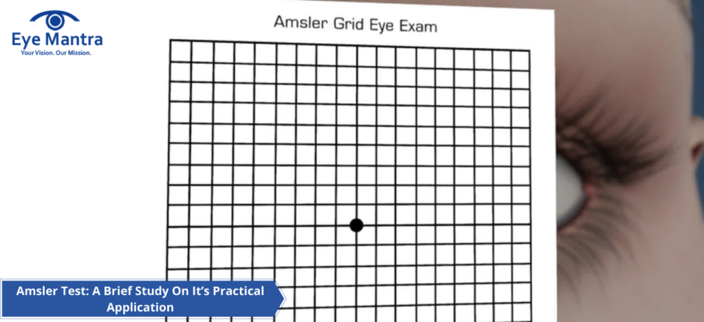
Amsler Grid
The Amsler Grid appears as a square-shaped grid that detects or monitors “metamorphopsia” or “scotoma” that involves the central visual field in several disorders of the macula and optic nerve head. It is mainly used as an inexpensive home monitoring device for the detection of metamorphopsia due to wet AMD at an initial stage.
The grid is named after Marc Amsler (1891-1968), who is a Swiss ophthalmologist.
Amsler grid appears like a piece of graph paper. It has a small dot in the center where you have to focus. While wearing your reading glasses, hold the grid at a distance enough to get most of the lines in focus. You can also monitor your vision at home with this chart if you are at risk for macular degeneration. But you cannot just rely on the chart for monitoring your eye illnesses, you should be regular in your visits to your doctor. A professional will always know more about the signs that you may miss.
Use of Amsler Test
The Amsler test or Amsler grid test can be useful in the following scenarios:
- Wet age-related macular degeneration.
- Central serous chorioretinopathy (CSCR)- It leads to a central scotoma that may be round or oval considering the shape of neurosensory retinal detachment.
- Metamorphopsia, caused by an epiretinal membrane and other vitreoretinal interface diseases.
- Acute macular neuroretinopathy- In this case, the scotoma is seen adjacent to fundus lesions. It becomes more prominent in multicolor imaging or near-infrared.
- Cystoid macular oedema- In this case, there is an increased difference between the photoreceptors due to macular oedema, and things may appear smaller than their actual size. This condition might result from diabetic maculopathy, retinal vein occlusion, intermediate uveitis, CNVM, and other diseases.
- Non-arteritic anterior ischemic optic neuropathy- In this case, the grid may show an altitudinal defect in the visual field.
- Central scotoma can be caused by hydroxychloroquine retinopathy. But the Amsler grid is not, however, recommended as a tool for hydroxychloroquine retinopathy.
- Pituitary tumor- Amsler grid demonstrates Bitemporal hemianopia
How To Use Amsler Test
- While conducting the test, make sure that the room lighting is normal as you use it for reading.
- Wear your normal glasses as you use them for reading.
- Amsler grid should be held approximately 14 to 16 inches from your eyes.
- Each eye should be tested separately (close one eye with your hand while testing the other one).
- While focusing on the center of the grid answer the following questions:
- Do you see any of the lines in the grid in a wavy, distorted or blurry manner?
- Is each box in the grid looking like a square and are all of them the same?
- Do you see any holes (missing areas) or dark areas in the grid?
- Are you able to see all corners and sides of the grid (while focusing on the center dot)?
After completing this with one eye, switch to the other eye, and repeat.
NOTE: If you experience any irregularities, report them to your optician immediately. Whichever areas you are not able to see properly in the Amsler grid, mark them. And bring the grid to the optician when you make a visit. Check your eyes with the Amsler grid as frequently as recommended by your optician or whenever you witness any irregularity.
Clinical Significance of Amsler Test
Amsler grid tool is significant for detecting or monitoring macular diseases.
Metamorphopsia- If some small squares appear to be widened than nearby small squares, then it may be denotive of Macropsia. There may appear a curve between the parallel lines in this area. The parallel lines seem to be drawn towards each other in the area of micropsia.
Scotoma- It is defined by Merriam-Webster dictionary as “a spot in the visual field in which the vision is either absent or deficient”. An area of the normal field of vision surrounds a scotoma.
To know more about it, you can easily visit our website Eyemantra. If you are looking for other services like cataract surgery, Retina surgery or Ocuploplasty you can simply ring at +91-9711115191. Even you can simply mail us on [email protected].
Medically Reviewed By

Senior Eye Surgeon | LASIK, SMILE & Cataract Specialist