Gonioscopy| All You Should Know About Goniscopy Test

GONIOSCOPY | ALL YOU SHOULD KNOW ABOUT GONISCOPY TEST
Gonioscopy is a test used by an ophthalmologist to check the drainage angle of the eye. This area exists in the front of your eye, i.e. between the iris and the cornea. A fluid called aqueous humour is naturally drained out by the eyes in this area. Gonioscopy is performed by your ophthalmologist to check if this part called the drainage
angle is functioning normally.
 
Under normal circumstances, the angle cannot be seen on exam. A special contact lens prism placed on the surface of the eye allows visualization of the angle and drainage system.

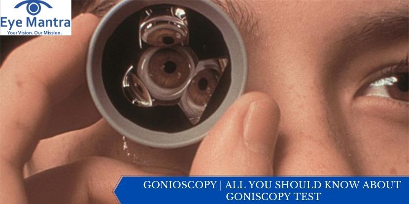
All About Gonioscopy Test

During a Gonioscopy exam, the patient is made to rest their head in the chin holder of a slit-lamp microscope, which is a special instrument that is used by an ophthalmologist to look in the patient’s eyes. At this point time during a Gonioscopy, the eyes of the patient are numbed with the use of eye drops.
 
Then, a special contact lens with mirrors is placed directly on the patient’s eyes by the ophthalmologist. The purpose of this is to shine a beam of light into the lens which would highlight the drainage angle. With the help of these lens mirrors, the ophthalmologist can see and analyze this part of the eye which is essentially very difficult to examine as it is around
the corner, inside the eye. The patient may feel the lens touching their eyelashes, however, the Gonioscopy exam is a
painless test throughout, from beginning till the end. The best part about the Gonioscopy test is that it is over in a matter of a few minutes.

Purpose of Gonioscopy Test 

Eyes are one of the most important senses of the human body, and to prevent dryness, they continuously make a liquid known as aqueous humour. However, it is important to maintain a cycle in the eye, where, the same amount of aqueous humour should be generated as much is excreted through a part of the eye known as the drainage angle. This process is necessary
 
 It maintains pressure in one’s eyes, which is known as intraocular pressure. In a case where the drainage angle is damaged or not functioning properly, a build-up of aqueous humour is caused in the eye. This leads to an increase of pressure in the eye, which ultimately damages the optic nerve, this is one of the main reasons why glaucoma is developed.
 
 The most common use of the Gonioscopy exam is to see if there are any signs of glaucoma in one’s eyes. The gonioscopy exam can help an ophthalmologist find out if the drainage angle of the patient is a bit too narrow for the
aqueous humour to flow properly, or in another case, blocked by part of the iris. There are many other conditions and ailments where the gonioscopy exam is performed on a patient, these include uveitis, eye trauma, tumors, etc.

Who Needs a Gonioscopy

By the time a person reaches the age of 40, one can detect early signs of vision changes and eye diseases. At this point of time in life, all people should get a baseline eye disease screening performed on them by an ophthalmologist. To search for signs of glaucoma, the ophthalmologist performs a gonioscopy exam on the patient to examine and analyze the drainage angle thoroughly, this involves its functioning as well as its appearance.


Gonioscopy and Glaucoma

Several people are termed as Glaucoma suspects, which means that even if they have normal pressure levels in the eyes, it is observed by their ophthalmologist that glaucoma can develop in their eyes. In such a case in addition to the gonioscopy exams performed by the ophthalmologist they also go under other glaucoma examinations regularly, to observe changes over time. Time is an important key when it comes to examining one’s eyesight. It is of extreme importance to have regular appointments scheduled with your ophthalmologists.
 
These regular eye check-ups and examinations can be the key to having a healthy vision.
Gonioscopy is a test performed by your doctor with a handheld goniolens. Some doctors refer to the lens as an “exam contact lens.” The doctor will barely touch the gonioscopy lens to the numbed cornea. The procedure is simple, quick, and does not hurt. It is similar to using mirrors in a periscope to see areas that are not otherwise visible to the naked eye.


Types of Goniolens or Gonioscope

The whole process of Gonioscopy varies according to which goniolens is used in the exam,
Some examples of goniolenses are as follows –

Koeppe Direct Goniolens

This lens is transparent, and the ophthalmologist places it on one’s cornea directly with some lubricating fluid, which helps him avoid any damage that can be caused to the surface of the eye. This goniolens’s exterior surface has a very steep curvature, which optically eliminates the total internal reflection and provides the ophthalmologist with a clear view of the iridocorneal angle also known as the drainage angle. However, this requires the patient to be laying down, hence it is impossible to be easily used with the help of a normal slit lamp in an optometric setting. However, a functioning microscope is an available option, when it comes to an ophthalmological setting.

Goldmann Indirect Goniolens

This lens is truncated in shape and it uses mirrors to reflect the light from the drainage angle towards the direction of the ophthalmologist. When used, the image of the drainage lens is roughly orthogonal to the back surface, which makes it very easy and reliable in making observations and magnifications with a slit lamp. The curved side of the lens is not rested on the cornea, but vaults around it, and the ophthalmologist uses lubricating fluid to fill the gap. On the sclera, rests the border of the front surface of the lens. This lens can be used with the patient sitting upright, and not laying down. Other mirrors of this device can also be used to analyze other parts of the eye, which include the retina and the ora serrate.

Zeiss indirect goniolens

This device functions using a similar technique as the Goldmann. However, this device uses prisms instead of mirrors. It has four symmetrical prisms which enable visualization of the drainage angle, in four parts of the eyes simultaneously, which makes it work well with a slit lamp. The most significant part of this device is that it has a smaller front, that rests on the cornea, where no lubricating fluid is required to fill the gap, and only the patient’s tear film allows for indentation gonioscopy, which helps the ophthalmologist perform further and more detailed diagnosis on the patient. 

Gonioscopy at Eye mantra 

Many optometrists perceive gonioscopy to be difficult to perform and shy away from performing it routinely. It is a learned skill that requires a great deal of experience. While there is a learning curve, with perseverance it can be mastered. Over time, the practitioner will learn to stabilize the gonioscopic image by making micro adjustments as the procedure is being performed. Proper technique is important, as undue pressure may open a narrow angle, giving a false clinical impression. Many eyes need to be observed to understand the subtle differences between the normal and abnormal angle. So it’s good to carefully choose the best possible staff who can facilitate such complex processes with ease. Therefore at Eye Mantra, here we have the most skilled doctors. 

The best way to treat your eyes is to visit your eye care professional and get your eyes checked regularly. He will be able to assess the best method of treatment for your eye ailment. Visit our website Eyemantra. To book an appointment call +91-9711115191. Or mail us at [email protected]. Our other services include Retina SurgerySpecs RemovalCataract Surgery, and many more.

Related Articles:

Myopia (Nearsightedness)

Dry Eye

What is Vision 20/20?


Make An Appointment

Free Tele-Consultation

Book Appointment or Video Consultation online with top eye doctors