
Contents
Worm In The Eye
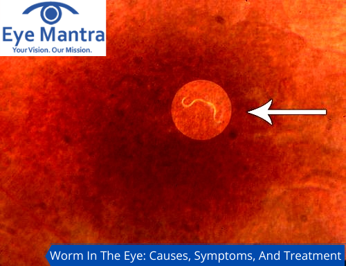
Diffuse Unilateral Subacute Neuroretinitis (DUSN) is a condition, where nematode (a type of eye worm) is present in the eye which can cause various eye problems. It may also cause temporary and permanent vision loss.
When larvae of a worm-like species, lodges in your eyes, then that worm larvae itself grows as a parasite in the eye, leading to vision problems. Eye worms are different from eye floaters (these are pieces of tissues that float in the fluid of the eye and sometimes resemble as worm-like species floating in the eye. These do not cause any harm to the eye but a sudden increase in their count is not a good sign. Whereas eye worms are actual parasites that lodge inside the eye and cause various conditions such as gnathostomiasis, loiasis, and onchocerciasis.
Some Examples of Eye Worms
- Gnathostoma spinigerium
- Loa loa
- Onchocerca volvulus
- Taenia solium
Symptoms of Eye Worms

Diffuse Unilateral Subacute Neuroretinitis has the following symptoms such as paracentral or central scotomas, early loss of vision, decreased vision loss in later stages. Deficits in the visual field and the presence of new floaters and severe pain. DUSN is usually seen as a worm in almost 20%-30% of patients suffering from worm eye diseases. There are various symptoms that can be helpful in determining the eye worms’ presence in the eye such as
- Presence of worm inside the eye
- Redness of the eye
- Severe pain
- Eye inflammation
- Retina scarring
- Watering of eyes and scarring of the retina
- Blurry vision, double vision, light sensitivity, loss of vision,
- Floaters in the eyes
- Discharge from eyes
Causes of Worm In The Eye
The causes of this condition are a helminthic infection with Toxocara Canis, Baylisascaris procyonis or ancylostoma caninum, or Gnathostoma spinigerium. The nematode cause damage through inflammatory, autoimmune and toxic mechanisms.
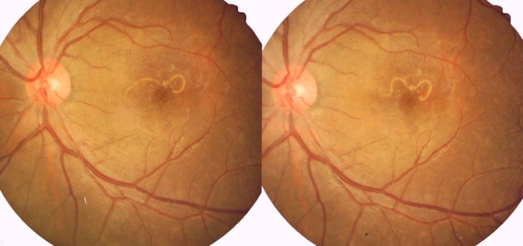
The outer retinal area is infected by local toxic effects and the inner retinal area undergoes diffuse toxic effects. The presence of worms is usually seen in the subretinal area and rarely seen in the inter-retinal area. DUSN is seen unilaterally, but rarely bilateral cases are seen. These worms cause peripheral loss of vision by inflammation in the retina.
The ingestion of nematode eggs takes place after they are shed by carriers such as dogs. Through blood, the nematode eggs travel over a period of many months and reside in the fundus region for almost 3 years. The unilateral loss of central vision is severe and insidious due to inflammation of the retina and degeneration of the retina. In DUSN, some changes were observed in the retina, subretinal space, optic nerve in DUSN.
Diagnosis of Eye Worm
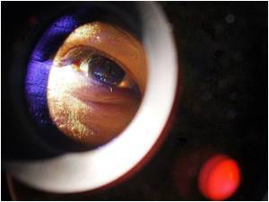
The worm inside the eye can be detected in the early stages. The appearance of the nematode inside the eye itself is the definitive diagnosis. Early diagnosis of the worms inside the eye is more effective than diagnosing them in later stages.
There are various diagnostic procedures that help in identifying the eye worms such as Fundus examination, electroretinography, Ocular coherence tomography, and scanning laser ophthalmoscopy. Laboratory test serum eosinophilia helps in diagnosing worms.
Eye Worm Treatment
DUSN is defined as Diffuse Unilateral Sub acute Neuroretinitis. This is a condition seen rarely in young people where a nematode is present in their sub-retinal area. The signs of this disease are differentiated into two parts, acute and end-stage manifestations.
In acute manifestation, the patients often have papillitis, also called optic neuritis, vitritis, and have decreased visual activity. Clusters of gray-white or yellow-white outer retinal lesions are seen. This helps in the localization of the nematode inside the eye.
During end-stage and if the worm is still off untreated, then the patient can develop optical atrophy, retinal arterial narrowing, and abnormal electroretinogram. Such conditions are often misinterpreted to be Unilateral retinitis pigmentosa.
Worms in the eye can be prevented if you take some precautions. you should avoid going to endemic areas and touching animals that can act as carriers of the etiological worms. It occurs in males than in females and even more frequently in young adults and children.
Systemic Treatment
Thiobendazole was used previously in treating this condition. Nowadays, albendazole which is given in doses of 400 mg/day for a month shows greater results and quick action. It has a few adverse effects such as stomach upset, dizziness, and rashes. Though it is not advisable for patients who are pregnant or lactating mothers as it can lead to teratogenic effects. Prednisone, given in 40mg – 60mg doses is the corticosteroid therapy recommended, then it is followed by taper over 2 or 4 weeks.
One can notice changes in the retinal structure corresponding to visual functioning through imaging modalities such as Ocular coherence tomography (OCT).
DUSN is treated with the help of strong medications in the early stages. However, during later stages, it can be treated with the help of surgery. The visualization of the worm in diffuse unilateral subacute neuroretinitis is a bit difficult because the worm often moves away from the light region. When a nematode is seen (in rare cases), it can be treated with photocoagulation. The use of anthelmintic drugs helps in the treatment of DUSN in the early stages. The anthelmintic therapy provides helps when the laser treatment does not kill the worm alone. Oral corticosteroids help in treating inflammatory responses around the eye.
Surgery For Worm In The Eye
Surgery is performed in severe cases when the worm is clearly visible. It is done with the help of high magnification biomicroscopy and fundus contact lenses.
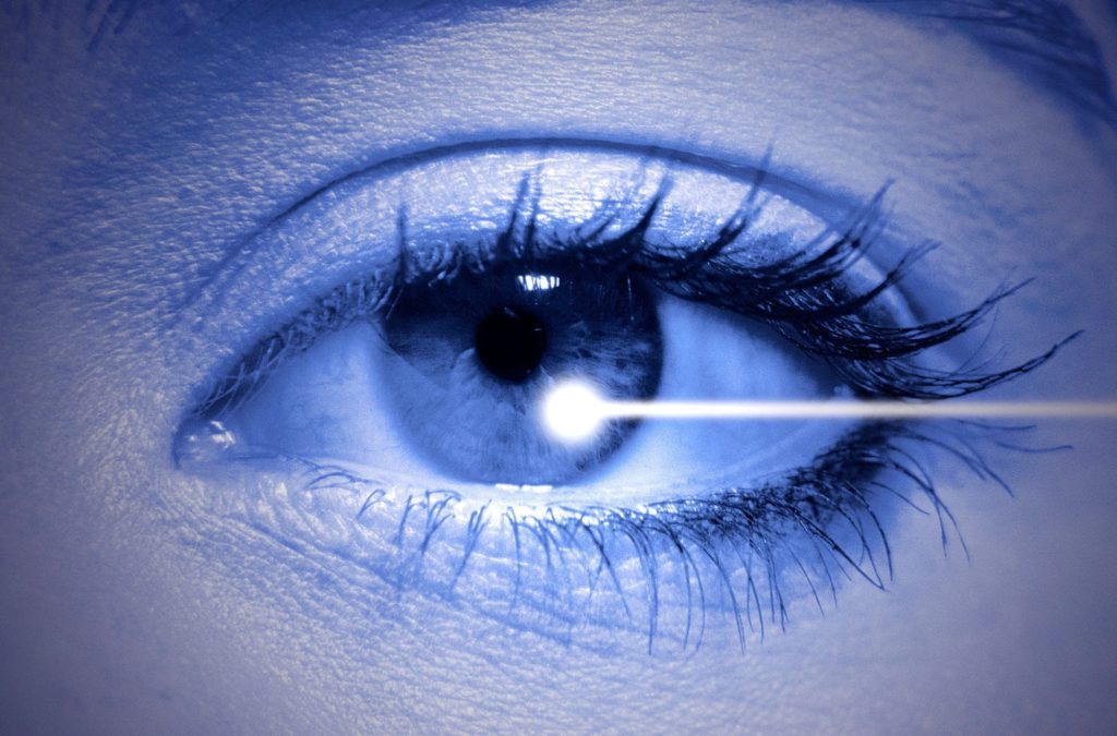
first of all, the worm is confided in a laser boundary through contiguous 200-micrometer spots. A laser of 200 mW power for 0.2 seconds duration is used for performing the surgery. The shriveling of the worm is seen when the burn is sufficiently intense. A single laser shot is sufficient to immobilize the worm at the head. A slit beam is used to prevent the movement of the nematode into delicate areas as the nematodes are photophobic in nature i.e, they move away from light.
Laser photocoagulation is a technique used in treating DUSN conditions after the worm is observed on a cluster of outer retinal lesions where the worm is supposedly present. Identification of worms is possible in 30% of affected cases.
During the early stages of infection, the treatment is done by using PSLP or laser by forming a laser grid in multiple spots of the retina in a faster time phase. Laser is used to preventing the movement of worms inside the eye. In a case where the worm is longer in size, then the laser shall first attack at the head end of the worm so that it prevents mobilization of the worms and makes it immobile. The speed of the laser prevents the movement of the worm inside the fovea.
Prognosis
The detection of disease in its early stages and treating them before it gets any worse can help in preventing loss of vision.
In later stages, the treatment can prevent the additional loss of vision. Any failure in nematode elimination in any of the cases can result in progressive chorioretinal destruction as well as sometimes severe and irreversible loss of vision. In later stages of DUSN, medical or surgical treatments do not improve visual acuity.
Complications of Surgery
The direct helminthic damage inside the eye is not reversible. Sometimes, photocoagulation can lead to hemorrhages, vascular occlusion and choroidal neovascularization, and epiretinal membrane formation. These changes can cause severe complications in the patients. After photocoagulation, the secondary inflammation gradually subsides after some time.
The best way to treat your eyes is to visit your eye care professional and get your eyes checked regularly. He will be able to assess the best method of treatment for your eye ailment.
Visit our website Eyemantra. To book an appointment call +91-9711115191. Or mail us at [email protected]. Our other services include Retina Surgery, Specs Removal, Cataract Surgery, and many more.
Related Links
Regular Retina Test: Purpose, Importance, and Risk
Medically Reviewed By

Senior Eye Surgeon | LASIK, SMILE & Cataract Specialist