
Imagine a small freckle in your eye – that’s a Choroidal Nevus for you. But is it just a harmless spot, or does it signify something more? As mysterious as it sounds, understanding the Choroidal Nevus is vital for anyone giving their eye health the attention it deserves. In this guide, we’ll demystify this tiny eye freckle, diving deep into its causes, symptoms, and the latest treatments available. Whether you’re a curious soul or someone keeping a vigilant eye on their vision (pun intended!), you’re about to unravel the intricacies of one of the most overlooked eye conditions. Let’s embark on this enlightening journey together!
Contents
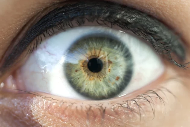
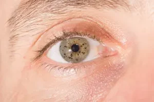
What is a Choroidal Nevus?
A Choroidal Nevus might sound like an enigmatic term, but think of it simply as a ‘mole’ or ‘freckle‘ inside your eye, specifically on the choroid layer, which is rich in blood vessels and lies between the retina and the white part of the eye, the sclera. Now, just as you have freckles or moles on your skin that are usually harmless, a Choroidal Nevus is typically benign too. However, its location within the eye makes it an area of interest for eye specialists. Compared to other eye conditions, a Choroidal Nevus is unique because it doesn’t typically affect vision or exhibit noticeable symptoms. Yet, it’s essential to monitor it, ensuring it remains harmless and doesn’t transform into something more serious. So, while it might be a tiny speck in the vast realm of eye health, understanding and keeping an eye on this ‘ocular freckle’ is crucial.
The Underlying Causes of Choroidal Nevus
 While the exact cause of a Choroidal Nevus remains a subject of ongoing research, several factors have been associated with its development. Here are some of the known contributors:
- Genetics: Just like skin moles, a predisposition to having a Choroidal Nevus can run in families. If close family members have it, you might be more susceptible.
- Ultraviolet (UV) Exposure: Extended exposure to UV rays, especially without protective eyewear, might increase the risk of developing an ocular nevus.
- Age Factor: The prevalence of Choroidal Nevus tends to increase with age. Older individuals are more likely to have this condition compared to younger ones.
- Eye Color: Interestingly, individuals with lighter eye colors, such as blue or green, may be more prone to developing a Choroidal Nevus than those with darker eyes.
- Hormonal Changes: Some studies suggest a link between hormonal shifts, especially during pregnancy, and the appearance or growth of nevi in the eye.
While these factors might increase the chances of having a Choroidal Nevus, it’s essential to note that the presence of one or more of these factors doesn’t guarantee its development. Regular eye check-ups remain the best way to detect and monitor any changes related to this condition.
Spotting the Symptoms: What to Look For
A Choroidal Nevus is often referred to as the “silent speck” of the eye world. Why? Because it predominantly remains asymptomatic. However, understanding potential signs associated with it is essential for early detection and monitoring:
- The irony with a Choroidal Nevus is that in most cases, there are no noticeable symptoms. It’s often discovered accidentally during routine eye exams.
- Visual Disturbances: In rare instances, if the nevus is located near the central vision area, it might cause slight visual disturbances or blurry spots in one’s vision.
- Visual Field Changes: A person might notice a small blind spot in their field of vision, especially if the nevus is larger or positioned near the optic nerve.
- Flashes or Floaters: Occasionally, individuals with a Choroidal Nevus might report seeing occasional flashes of light or floating specks in their vision.
- Changes in Appearance: Any transformation in the size, shape, or color of the nevus should be noted, though this is typically observed by an ophthalmologist rather than the individual.
Given the typically silent nature of a Choroidal Nevus, the emphasis on regular eye examinations cannot be overstated. It’s these routine check-ups that ensure its early detection and timely intervention if any changes are observed.
The Link Between Choroidal Nevus and Melanoma
A Choroidal Nevus, in many ways, can be compared to a mole on the skin. Just as most moles are benign and harmless, so too are most Choroidal Nevi. However, just as there’s a possibility for certain moles to turn malignant. Choroidal Nevus can progress to Choroidal Melanoma—a type of eye cancer.
Here’s what you should know:
- Frequency of Transformation: While the progression of a Choroidal Nevus to Melanoma is relatively rare, it’s a transformation that can’t be entirely ruled out. Estimates suggest that approximately 1 in every 8,000-10,000 nevi might undergo this change.
- Risk Factors: Certain characteristics of a nevus can make its transformation more likely. These include greater thickness, the presence of orange pigment, proximity to the optic nerve, and the presence of subretinal fluid.
- Importance of Monitoring: Due to the potential for malignant transformation, it’s essential to keep a watchful eye on any diagnosed Choroidal Nevus. Regular eye exams and imaging tests help track any changes in the size, shape, or appearance of the nevus.
- Early Detection: If detected early, Choroidal Melanoma is treatable. However, like all cancers, early diagnosis is crucial to successful treatment and positive outcomes. This further underscores the significance of regular check-ups and monitoring of a Choroidal Nevus.
It’s worth noting that the vast majority of Choroidal Nevi remain benign throughout a person’s life. Yet, given the gravity of a potential melanoma diagnosis, vigilance is always advised. If ever in doubt, or if you observe changes in your vision, it’s essential to consult with an ophthalmologist.
Diagnostic Procedures for Choroidal Nevus
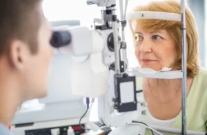 Diagnosing a Choroidal Nevus requires specific tools and methods that eye professionals employ to ensure accurate detection and assessment. Here’s how the process typically unfolds:
- Comprehensive Eye Exam: The journey starts with a thorough eye exam. Using a tool called an ophthalmoscope, the ophthalmologist will inspect the back of your eye, searching for any abnormal growth or discoloration.
- Fundus Photography: This technique captures detailed images of the retina, optic nerve, and blood vessels. It can help in documenting the size, shape, and location of a nevus, allowing doctors to monitor any changes over time.
- Fluorescein Angiography: By injecting a fluorescent dye into the bloodstream and taking images as the dye passes through the retinal vessels, this test helps in examining the nevus’s blood flow and structure.
- Optical Coherence Tomography (OCT): Using light waves to capture cross-section pictures of the retina, OCT provides a clearer view of the nevus’s thickness and its relation to the different layers of the retina.
- B-scan Ultrasonography: This test uses sound waves to create an image of the inside of the eye. It’s especially useful when the nevus is located in an area that’s hard to visualize with traditional tools or when it’s positioned deeper within the eye.
Early and accurate diagnosis of a Choroidal Nevus is crucial for effective management and monitoring. If you ever notice any changes in your vision or if Choroidal Nevus runs in your family, don’t hesitate to schedule a comprehensive eye examination.
Modern Treatment Approaches for Choroidal Nevus
In the landscape of eye health, the management of a Choroidal Nevus has witnessed considerable advancements. The treatment approach primarily depends on the size, location, and associated symptoms or risks of the nevus. Here’s an overview of the modern treatment options available:
Observation
- Description: Often, if the Choroidal Nevus is small and shows no signs of growth or risk of progression to melanoma, the best approach is observation.
- Effectiveness: It’s a safe way to ensure that any changes or unusual developments are detected early.
- Potential Side Effects: There are no side effects to this approach, but the key is regular check-ups to ensure timely detection of any changes.
Laser Photocoagulation
- Description: This involves using a laser to treat the nevus. The laser energy creates a thermal effect, causing coagulation.
- Effectiveness: It’s primarily used for nevi that show signs of growth or those situated close to the retina to prevent complications.
- Potential Side Effects: Mild pain, temporary vision disturbances, or minor bleeding.
Photodynamic Therapy (PDT)
- Description: A light-sensitive drug is introduced, which gets absorbed by the nevus. Later, a specific wavelength of light activates this drug, destroying the abnormal cells.
- Effectiveness: It’s beneficial for treating nevi that pose a potential threat but are not yet malignant.
- Potential Side Effects: Light sensitivity for a short duration, localized swelling, or minor pain.
Radiotherapy
- Description: In cases where there’s a high risk of the nevus transforming into melanoma, localized radiation might be considered.
- Effectiveness: It prevents the growth of abnormal cells, thus mitigating the risk of malignancy.
- Potential Side Effects: Temporary or permanent vision disturbances, dry eyes, or mild inflammation.
Surgery
- Description: In rare cases, if the nevus poses a significant risk or causes vision impairment, it might be surgically removed.
- Effectiveness: Direct removal eliminates the risk associated with that particular nevus.
- Potential Side Effects: As with any surgery, there’s a risk of infection, bleeding, or post-operative complications.
It’s important to note that the treatment approach is personalized. Not all patients with a Choroidal Nevus require intervention.
Conclusion
In conclusion, with modern medical advancements, managing and treating this condition has become more precise and patient-centric. Remember, your vision is a priceless asset, and it deserves the best care and attention. If you find yourself grappling with retina-related concerns, know that help is just around the corner. At EyeMantra, our dedicated team of experts is equipped to guide you through your journey to optimal eye health.
If you’re experiencing retina-related problems, Retina Surgery at EyeMantra can help: Book your free appointment now at 9711116605
Medically Reviewed By

Senior Eye Surgeon | LASIK, SMILE & Cataract Specialist