A general layman perception about eye testing is that is only for checking the power of the glasses we need. However, there is much more to a full eye examination than visual acuity tests. Visual acuity tests involve the assessment of the reading capacity of eyes from a given distance. The patient is asked to read out the fonts of different sizes while covering one eye at a time, and with both eyes open.
Based on how conveniently one can read the alphabets and characters, the optometrist keeps changing the lens combinations until appropriate power is identified. This is the most basic and manual method of evaluating visual acuity, which is also often practised by non-specialists and opticians locally. Ophthalmologists also supplement it with a mechanical test for approximation of eye power.
It is very important to maintain and follow up with your regular eye check-ups. Some people take this very lightly without realizing the outcomes. Eye examinations help us to find the problem or disease that is creating its space in our eyes. If we examine and treat it in its early phase we can get our healthy eye back whereas if we just sit back and do nothing it can worsen the situations of our eye. There are several cases in which people go blind just because they didn’t go for treatment at the early stage of eye problems.
Contents
When To Go for Eye Exam
If you have any of these symptoms make sure you for go an eye exam:
- Red, dry and itchy eyes.
- If you have diabetes or any other problem affecting your eyes.
- You have flashes and you see a black dot floating i.e. floaters in your vision.
- Difficulty seeing in dim lights or at night.
- If you have a severe headache or eye strain after spending several hours on the screen.
- You get motion sick, dizzy, or have trouble following a moving target.
- You are unable to read the newspaper properly i.e. you have blurry visions.
- Unable to see things clearly after having some head injury.
What is Retina?
The retina is the back screen of our eye where all the images are projected. It is the retina that is responsible for our vision. The retinal disorder leads to disrupted visions formed in our eyes. If our retinal nerves are damaged, then the retina cannot send proper signals to the brain, leading to blurry or unclear images. There are many retinal conditions and diseases, but most of them can be treated if and when they’re detected early. Several eye exams can detect problems with the retina early to help you avoid serious diseases and complications. It is recommended that you visit your eye doctor annually to keep your eyes healthy. You may not see any symptoms in such cases. You may not even feel any pain and may continue to see sharply even though your retina is impaired. Problems in the retina can be a symptom of the following diseases:
Retinal Diseases
A common retinal problem experienced by people is retinal detachment. The middle of our eye is filled with a clear gel that helps in image formation. Due to some injury or disease, tiny clumps are formed in this gel, resulting in shadows on our retina. This gel may shrink, pulling on the retina bit. That could cause us to see flashes. These are common symptoms of this disease and do not generally do us any harm. If the clear gel moves too far away from the retina, it can tear the retina, leading the fluid to leak into the tissue, leading to retinal detachment. Therefore, your retina is not able to send proper signals to the brain the vision will become blurry. This may also lead to blindness quickly if left untreated.
Floaters
It causes spots in your vision. They can be age-related, but they can also occur in cases of severe near-sightedness. However, floaters can be formed due to a torn retina. If the tear isn’t repaired at an early stage, then it can lead to retinal detachment. This is due to fluid that forms behind the retina, leading to its separation from the eyes.
It is an age-related condition of the retina that causes vision loss. It is common in individuals and people over the age of 55 are prone to this disease. The symptoms involve blurry vision, warped straight lines, or difficulty focusing on fine details. Blind spots can be developed as the condition worsens. There are several treatments for this condition. Make sure you don’t wait for the condition to get worse.
Diabetic Eye Disease
It damages the blood vessels in your retina. Over time, it leads to vision loss if not controlled on time. Major symptoms can be blurry vision, double vision, floaters, eye strain, flashing lights, or rings.
Laser surgery is has been recommended being the most efficient treatment for a person suffering from diabetic eye disease. It is important to know that increases in the risk of diabetes may also lead to glaucoma and cataracts.
Retinal Detachment
Retinal detachment occurs when too much fluid is generated behind the retina, causing separation. However, there are other risk factors that increase the chances of retinal detachment such as:
- Extreme near-sightedness
- Previously a case of retinal detachment in the other eye
- Genetic predisposition
- Previous cataract surgery
- Presence of any other eye disorders
- Eye injury or head trauma
The presence of floaters indicates the beginning of retinal detachment. There may also be flashes in the eye. If it is not treated quickly, it can cause permanent vision loss. If you suddenly begin to see floaters in your vision, visit a doctor immediately. Other symptoms include a decrease in vision or blurry visions. You can save yourself from permanent vision loss by treating the cause of the disease by its roots.
Several eye diseases may only harm your eyes at night like you may have difficulty while driving at night whereas there are some diseases that may affect your eyes during the day like you get sensitivity to light or you see black spots everywhere in your vision. You need to be very vigilant about your eye diseases and consult a doctor if you have any kind of problem in your eyes.
What is a Retinal Examination?
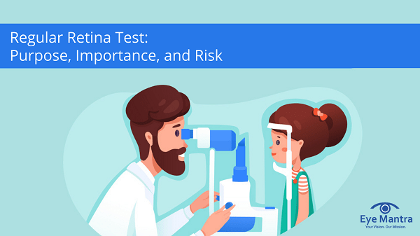
A retina examination helps your eye examiner to find the problems in the back of your eye. Your eye doctor will use a bright light and look through a microscope to examine the optic nerve, retina, and blood vessels, also known as slit-lamp examination. The process is comprised of four parts.
Retinal imaging takes a digital picture of the back of your eye. It shows the retina (where light and images hit), the optic disk (a spot on the retina that holds the optic nerve, which sends information to the brain), and blood vessels. This helps your optometrist or ophthalmologist find certain diseases and check the health of your eyes.
Doctors have long used a tool called an ophthalmoscope to look at the back of your eye. Retinal imaging allows doctors to get a much wider digital view of the retina. It doesn’t replace a regular eye exam or regular dilation,, but adds another layer of precision to it.
- Dilating the Eyes –firstly the doctor will put some eye drops in your eyes. This helps the pupil to get widen so that the eyes can be examined in detail.
- Tonometry Test – Tonometry measures the pressure inside the eye. This helps your ophthalmologist look for signs of glaucoma. A puff of air is blown directly into your eye.
- Visual Field Test –this test is done to see the focal length of your vision. One eye is tested at a time. You will be asked to focus on some image and then the machine automatically gets the results.
- Visual Acuity Test – This test involves reading an eye chart. In which the size of the letters gradually decreases as you go down. This is the most common eye test to check your vision.
Who should go for a Retinal Test?
Diabetes: when your body starts to lose control over this disease and you get vision problems. This disease is one of the major reasons for eye problems. It is advised to get an eye examination done at the beginning of the disease only, so as to avoid any further complications.
Macular degeneration: The central part of your retina (the macula) gets worse with age. You may have blurry vision and difficulty in focusing on some object. There are two kinds of macular degeneration: wet and dry.
Dry macular degeneration is by far the most common form of this disease (up to 90% of the cases). It happens when blood vessels under the retina become thin causing trouble seeing the object clearly.
Abnormal blood vessels growing under the retina cause wet macular degeneration. Vision loss is usually fast.
Retinal imaging is very important in finding this type of macular degeneration.
Glaucoma: This disease damages your optic nerve (located in the retina) and may cause vision loss. It typically happens when fluid builds up in the front of your eye. It can cause blindness but it normally progresses slowly and can be treated with special eye drops to lower the pressure caused by the fluid
To know more about it, you can easily visit our website Eyemantra . If you are looking for other services like cataract surgery, Retina surgery or Ocuploplasty you can simply ring at +91-9711115191. Even you can simply mail us on [email protected].
Related Articles:
Quick Home Remedies for Pink Eye
Computer Eye Strain Relief


