
Contents
What Is Coloboma?
It is a birth defect found in children where there is an absence of tissue in any part of the eye. After fertilization in the womb, the eyes are the first organ that is developed in the embryo. It starts to develop from the optical vesicle. The lower end of the optical vesicle consists of a groove called the choroidal fissure. In this groove, the entry of blood vessels takes place for the eyes. The fissure normally closes by the seventh week of pregnancy. In some embryos, it fails to close this leads to the formation of a gap or space where tissue remains absent and the condition is termed as “coloboma”.
It can occur in one eye or both eyes and can affect them differently. The disease affects around 1 in 10,000 children and the Indian subcontinent has a higher frequency of newborns affected with this eye disease. In some children, the condition may be adverse and would lead to vision loss but in some, it may not affect vision and may remain unnoticed for a long period of time. Coloboma is inheritable and can be transferred through the autosomal genes and in very rare cases through the X- linked inheritance. It is also associated with other eye diseases and can lead to complications in the eyes of the newborn.
Types of Coloboma
According to the parts of the eye which has an absence of tissue, Coloboma can be distinguished into different types.
Eyelid Coloboma
It is an uncommon, unilateral or bilateral, partial or full-thickness eyelid defect in which a part or whole eyelid is missing in the child. It is caused due to the failure of fusion of the mesodermal lid folds. It can be linked to various other ocular or systemic anomalies. If eyelid coloboma is left untreated for a long period of time then it can lead to complications like corneal leukoma, symblepharon, and amblyopia.
There have been studies that suggest that this tyoe is linked with intrauterine factors including amniotic bands, abnormal fetoplacental circulation, or radiation. They are also found in association with the facial clefts, Goldenhar syndrome, Treacher Collins syndrome, charge syndrome, or frontonasal dysplasia.
Eyelid reconstruction is important in the patients depending upon the size of the defect and the presence of corneal exposure. Surgery should be done as soon as possible to avoid corneal lesions. In children having mild conditions, surgery can wait till the age of 3-4years. Eyelid coloboma poses a potential threat to vision loss in newborns and should be treated as early as possible to avoid other complications in the eyes.
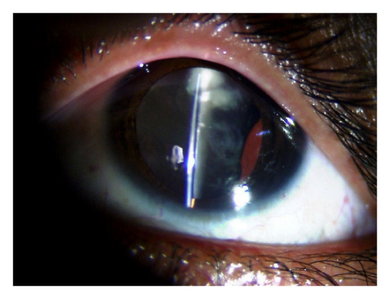
Lens Coloboma
It is defined as the anomaly of lens shape and is mainly the result of defects in zonules and ciliary bodies during ocular development usually unilaterally. If the zonule of the ciliary body fails to develop, the tension in the eyes is released and the contract segmentally a notch. It appears in combination with other defects including iris, choroid, or optic disc coloboma.
The patient having this has a higher risk of developing a detached retina and cataract. Lens coloboma is also linked to other diseases including Marfan syndrome and Marshall syndrome. Treatment of this disease usually involves surgical methods and phacoemulsification with capsular tensor ring and intraocular lens implantation.
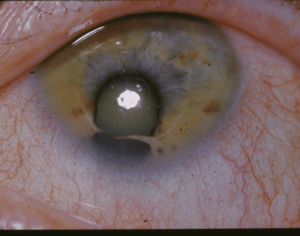
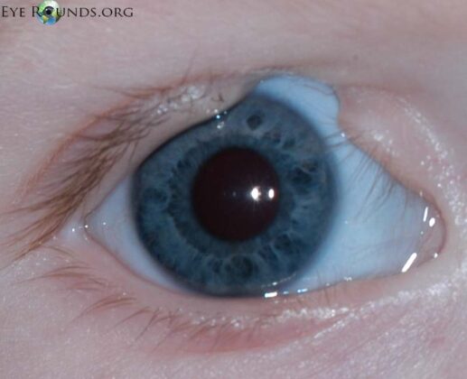
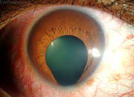
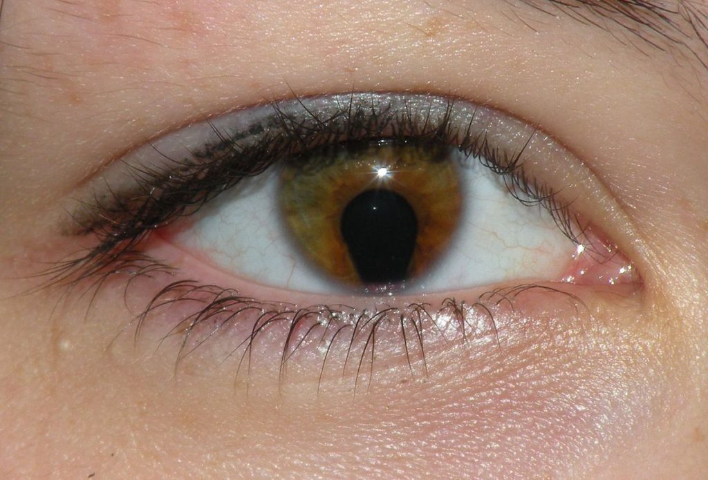
Iris Coloboma
The iris present in the eyes is known to perform the function of controlling the amount of light entering the eye by adjusting the size of the pupil. When there is a defect in the iris or there is the presence of iris coloboma then the iris is not able to perform its natural function. This causes glare-sensitive eyes in the patients having iris coloboma as the scattered light is able to reach the eye due to enlarged pupils.
Traumatic iris coloboma is an acquired condition that occurs due to the result of an accident when the iris is ruptured or may occur after glaucoma surgery. Typical iris coloboma generally occurs in the lower and inner quadrant of the iris, giving the pupil a keyhole appearance. It can also be associated with other ocular disorders including ciliary body coloboma, choroid-retinal coloboma, optic nerve coloboma, and small eyeball. It is the most common type of coloboma that occurs in infants.
Macular Coloboma
The macula is the part of the eye that is responsible for the central vision and is a part of the retina of the eye. It is a rare, non-syndromic developmental defect of the eye that can be characterized by well-circumscribed, oval or rounded, unilateral, atrophic lesions present in various sizes showing a rudimentary or absence of retina, choroid, and sclera located at the macula part of the eye. It leads to decreased vision or complete vision loss. It is present in isolated- conditions and in rare cases can be found in association with autosomal dominant and other inheritance patterns including down syndrome, and skeletal or renal disorders. A rare atypical variant of macular coloboma is bilateral and symmetrical.
Optic Nerve
It is a congenital eye abnormality in which the optic nerve is incompletely formed. The condition may occur in one or both eyes. It may occur spontaneously or can be inherited from parents due to mutations in various genes or can be associated with other genetic conditions including CHARGE syndrome.
Optic nerve coloboma is associated with autosomal dominant inheritance of the mutated PAX6 gene which is responsible for forming tissues and organs in the developing embryo. As the condition is present from birth there may be a delay in the diagnosis as the condition is present inside the eyes and would not be visible in a simple inspection. Vision can be impaired based on the seriousness of the condition. Retinal detachment can occur in optic nerve coloboma.
Uveal Coloboma
The coloboma of the uveal is found in association with other ocular anomalies including systemic diseases. In this, there is the involvement of a complete keyhole-looking iris, oval pupil, heavily pigmented iris, ciliary body, retina, lens, choroid, and the optic nerve. Uveal coloboma is linked to autosomal dominant patterns along with homozygous mutations.
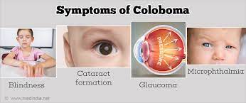
Symptoms of Coloboma
- The symptoms are generally seen after the birth of the newborn. The eyelid coloboma would be easily visible due to the presence of a notch or defect in the eyelid of the child. If the eyelid coloboma is large then it can lead to scarring and visual impairment in the child.
- The iris coloboma can be recognized by the keyhole-like or cat-eye-like appearance of the pupil. In some cases, there is duplication of images due to the small iris being not attached to the pupil of the eye. This causes blurring of vision in children having iris coloboma.
- The lens coloboma can also be recognized easily as there are high chances of it occurring along with other ocular anomalies.
- The children having coloboma of the optic nerve and macula would suffer from vision problems, they may also remain asymptomatic throughout their life but proper diagnosis can help detect the eye condition present in them. In some cases, the defect remains unnoticed in the early years and suddenly appears in form of vision loss in adulthood due to retinal detachment that occurs over time.
- Increased sensitivity to light can be seen in children having this disease.
- In children having coloboma which is associated with other genetic disorders then symptoms of that particular syndrome would be visible. There are other eye problems that are linked with coloboma including cataract formation, glaucoma, and subluxation of the lens.
Causes of Coloboma
Genetic
Coloboma can be caused due to genetic inheritance or may occur randomly in any embryo. In genetically inherited coloboma the pedigree that it follows may be autosomal dominant, autosomal recessive and in rare cases, it may be X-linked inheritance. PAX6 is associated with coloboma. It is a gene which in normal conditions is responsible for the development and formation of tissues and organs in the embryo. Any mutation in this gene causes the improper formation of the tissues of the developing embryo. It can also occur in association with another genetic syndrome like CHARGE.
CHARGE is an acronym for “Coloboma of the eye, Heart defects, Atresia of the choanae, Retardation of growth and development, Genital abnormalities, and Ear abnormalities”. This syndrome is caused due to mutation of the CHD7 gene present on chromosome 8 and is a life-threatening syndrome.
Environmental Factors
There are other environmental factors that also play a major role in causing this in children. A pregnant woman who is an alcohol addict or drinks alcohol during her pregnancy period tends to have a child born with the disease and causes fetal alcohol syndrome in babies.
Deficiency of vitamin A in mothers also causes risk of having a child born with coloboma. Intrauterine infections caused due to toxoplasma can also lead to this disease formation in the baby. Drugs like thalidomide or mycophenolate can also contribute to the formation of coloboma in the embryo.
Treatment
The treatment of coloboma depends on the type of it coloboma present in the children.
Eyelid Coloboma Treatment
To treat eyelid coloboma first detailed history of family, drugs taken during pregnancy, and complete ophthalmic exam is conducted. Initially, a visual acuity test is performed along with various other tests to determine any association of disease with other ocular anomalies and genetic syndrome.
Eyelid reconstruction is done in infants having coloboma of the eyelid. Defects up to 25% can be closed directly, defects ranging between 25-50% are repaired by direct closure with canthotomy and cantholysis or by tenzel’s semicircular flap. Defects more than 50% are treated using Tenzel’s semicircular flap, Cutler-Beard technique, or Mustarde rotational flap is used.
Lens Coloboma Treatment
The initial step is to obtain detailed imaging of the lens, zonulae, and ciliary body. Scheimpflug images are used to obtain more accurate and detailed images. Then surgery is performed by the phacoemulsification and intraocular lens placement. The surgical option is very challenging and can lead to capsular fornix aspiration, zonular dialysis extension, vitreous herniation, and closure of the capsular opening.
To reduce the surgical complication implantation of a capsular tension ring is done to prevent capsular bag deformity and avoid capsular collapse. Significant improvement in vision can be observed after the surgery. This method of surgery is considered to be safe and effective to treat coloboma of the lens.
Iris Coloboma Treatment
There are many treatment options available in coloboma of iris depending upon their severity. Blue-blocking filtered eyeglasses are prescribed to reduce contrast vision. Prosthetic contact lenses are used to reduce glare and mask the iris. The contact lenses mimic the natural iris pattern and create a new aperture for the eye through which light can enter.
Pupil occluding lenses are the lenses that are used to prevent ghost or double vision. Surgery performed on the iris can be done in order to implant a custom-made lens on the iris or a foldable artificial iris lens can be used. In implantation, the whole iris can be implanted or only the missing part of the iris can be implanted.
Optic Nerve Treatment
No particular treatment option is available to treat coloboma of the optic nerve as optic nerve one damaged cannot be transplanted or treated by surgery. This kind of coloboma is treated with the help of low vision aids which can be helpful to some people.
Uveal Coloboma Treatment
No particular treatment is available to cure uveal coloboma fully. Patients with this type are usually prescribed corrective contact lenses so that amblyopia does not develop in early childhood. Using low vision aids is also helpful in this condition.
Living With Coloboma
Coloboma cannot be fully treated or cured. Even after surgery, there are chances of having poor vision or the development of any other ocular anomalies. Often genetic counseling can help the parents and patients in being aware of the disease. The children having it which is associated with genetic syndrome need lots of love and care so that they don’t develop an inferiority complex about their syndrome. The chances of having this disease are less but it is essential for patients to brace for the vision problems related to the disease in the future.
Medically Reviewed By

Senior Eye Surgeon | LASIK, SMILE & Cataract Specialist