
Contents
How Can Photos Help Detect An Eye Illness?
Most of us take photos to capture the beautiful and crucial moments of our lives. But these photos can tell a lot more than just a moment. Photos nowadays are proving effective for detecting eye illness as cameras are able to do an early-stage diagnosis.
Illustrating with an example- A few years back Tara Taylor posted a picture with her 3-year-old daughter on a social media platform. Some of her friends informed Taylor that the white glow in her daughter’s eye could indicate an eye illness. Eventually, her daughter was diagnosed with a rare eye disease that can lead to vision loss. But due to early detection of the eye illness, her vision was saved.
After that, this trend gained popularity. People started clicking their pictures and shared them on social media to reach a wide audience. Looking closely at the pictures of children can actually give a clue about an existing eye illness.
When the flash of the camera lights up the blood-rich retina a red reflex is produced. If both eyes are focused on the camera and the color of the reflex reflects as red then it can be read as a good sign. It indicates that the retinas of both eyes are healthy and are not obstructed.
Flash photographs can also detect the most common early sign of retinoblastoma i.e., white pupil.
Signs of Eye Illness Captured By Camera
- One red eye– When there is no red-eye or one eye appears dimmer than the other eye, it can be an indication of Strabismus. It can be understood as a condition where both the eyes do not look or focus in the same direction. Strabismus can be treated by glass eye, covering the strong eye with an eye patch, or even an eye muscle surgery. Strabismus can be corrected if detected and treated early.
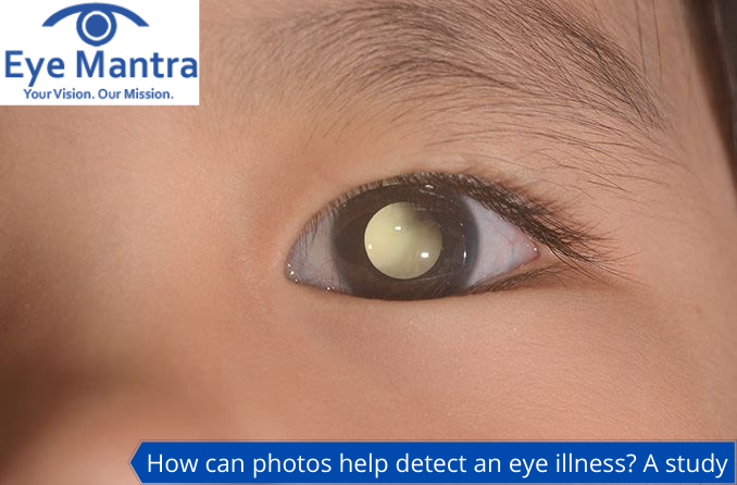
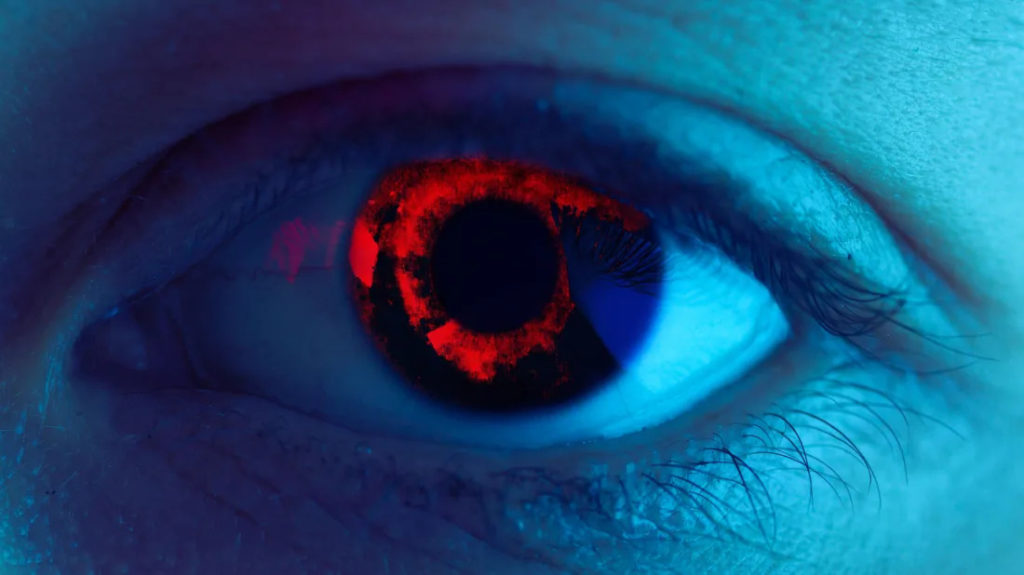
- White pupil– White pupil should be seen as an early warning. The medical term for this is leukocoria. If the pupil appears white, it may indicate several serious eye disorders such as retinal detachment, cataracts, or infection inside the eye. The pupil at this stage can be described as “a glow”, “milky”, “transparent”, “crescent moon” etc. It can also be understood as an early-stage sign of retinoblastoma. Retinoblastoma is a very rare eye cancer and can be cured 90% of the time.
- Yellow reflex– This is a disease that causes blood vessels inside the retina of the eye to become twisted and leaky. It causes a block in the retina leading to vision loss or even retinal detachment. The yellow reflex can be caused due to this disease.
Also Read:
What Are Scleral Lenses | Its Uses, Types and Facts
Serious Eye Disorders: White, Yellow, And Black Flags
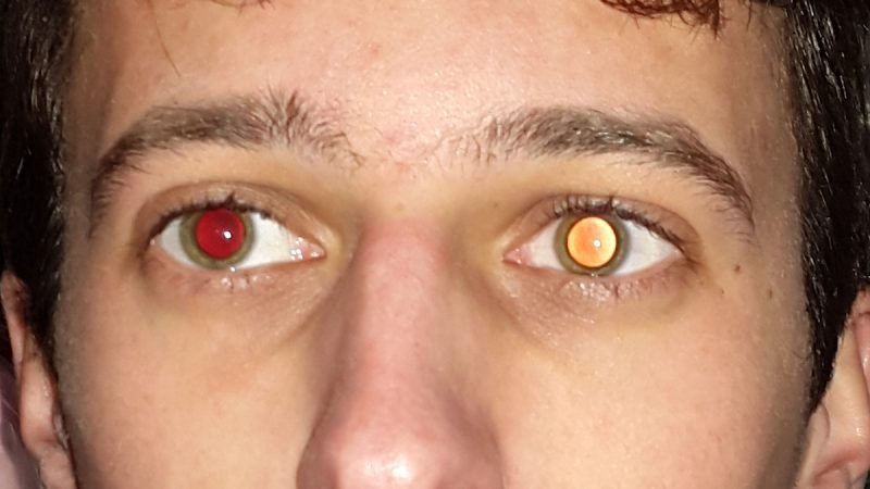
Abnormal red reflexes can indicate more serious eye disorders.
- Asymmetrical reflex: When only one eye appears as red and the other one does not or it appears dimmer than the former one, then it might be an indicator of strabismus. Strabismus is the misalignment of the eye where both the eye do not focus in the same direction. Strabismus can be treated with eyeglasses, prisms, patching, or blurring of the strong eye, and the last option is eye muscle surgery. Earlier detection and treatment of strabismus can deliver excellent results.
- Most of the pupil is covered by the White reflex which is also known as leukocoria. It may point to retinoblastoma which is an extremely rare childhood eye cancer. “90-95% of the time, this disease can be cured by early detection and treatment”.
- Yellow reflex: It may indicate Coat’s Disease, which happens when blood vessels of the eyes that provide blood and oxygen to the retina become twisty and leaky and create a blockage in the retina that can lead to vision loss or retinal detachment. It mainly occurs in boys who are below the age of 10. It affects one eye usually. Coat’s Disease can be treated with laser surgery, cryotherapy, surgery, etc.
People may find it hard to distinguish Coat’s Disease from retinoblastoma through photographs because the reflexes of white and yellow look quite similar.
Red-Eye Effect
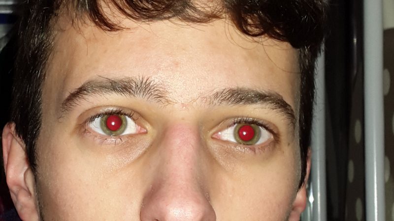
Abnormal Red eyes in photos can help detect eye problems and could provide potentially sight-saving information about the eyes. The red-eye effect is captured in dark or dim light. It occurs when the person is looking directly into the camera and the flash is on when the background light is dim.
The red-eye effect from photos is very common and mild. This effect occurs when the camera flash gets reflected from the retina. When light enters the eyes causing the pupil is to widen to allow light to reach the retina which is the inner layer of the eye. Retina receives the light signals and sends them brain which forms the image.
In the case of photographs, the flashlight is not absorbed completely by the retina and is reflected back creating the red-eye effect. It illuminates the blood supply in the retina producing the red color you see in the pictures.
PhotoRED Technique
The photoRED technique can be used for detecting the red-eye reflex. The camera is used as a diagnostic tool, in this technique.
- Smartphone flash is not reliable so use a regular camera
- Turn the lights dim so that the camera uses auto-flash.
- Make sure any light source like a table lamp is behind your child so that it does not reflect in the eye.
- Ensure that your camera’s red-eye reduction feature is turned OFF. The red-eye reduction symbol is usually represented as a diagonal line through an eye.
- Stand about four meters away from your child and zoom in on the camera to capture your child’s entire head.
- Take multiple photographs from different angles.
- Go through each and every photograph for abnormalities such as a white reflex, no red reflex, or reflexes that do not look similar in both eyes.
Parents should be vigilant about their child’s eyes because even the most dangerous eye diseases don’t cause children pain or visual impairment during the early stages.
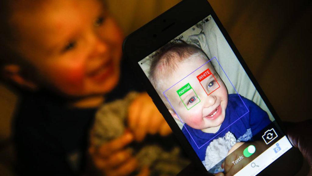
In most eye illness cases, one of the eyes of the child is working. Therefore, it becomes even harder to detect the disease. There are 90% of cases of retinoblastoma which have been detected by parents through photographs. In 2014 an app was developed for the detection of such abnormalities but this app cannot distinguish between the white and the yellow reflex. The name of the app is the White Eye Detector.
There are ongoing researches to develop software that can automatically detect leukocoria and other abnormalities in photographs. Scientists have even developed an app to scan through children’s eyes and detect if there is any kind of eye illness.
When To See Doctor?
In case you are successful in detecting any such illness, then get your child evaluated as soon as possible by a pediatric ophthalmologist. Doctors advise the parent to conduct the red-eye test every month to keep track of any such abnormality. Early detection and treatment of any such disease have higher chances of recovery.
The difference between the red-eye reflex should be the most cautious one. Parents should carefully analyze the photographs for any such conditions.
As we have progressed further, modern technology has removed the red-eye issue from the cameras. Modern cameras have a new feature known as the red-eye reduction feature. This red-eye deduction feature restraints the camera’s detection of the white pupil.
To know more about it, you can easily visit our website Eyemantra. If you are looking for other services like cataract surgery, Retina surgery or Ocuploplasty you can simply ring at +91-9711115191. Even you can simply mail us on [email protected].
Medically Reviewed By

Senior Eye Surgeon | LASIK, SMILE & Cataract Specialist